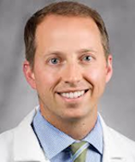
Brian Hinds, M.D.
· HS Clinical Associate ProfessorUniversity of California, San Diego · Dermatology
Active 2010–2026
About
Brian Hinds, M.D., is a Clinical Professor of Dermatology at UCSD. His research focuses on various aspects of dermatology, including immune signaling pathways, cutaneous malignancies, infectious mucocutaneous eruptions, and immune-related skin toxicities. Dr. Hinds has contributed to understanding the dual dysregulation of TNF/interferon signaling and the role of classical monocytes in reactive infectious mucocutaneous eruptions, as well as investigating the immune mechanisms underlying conditions such as Sweet syndrome and cutaneous T-cell lymphoma. His work also encompasses case reports and studies on skin manifestations related to infections, neoplasms, and drug reactions, demonstrating a broad engagement with both clinical and translational dermatology.
Research topics
- Medicine
- Dermatology
- Internal medicine
- Pathology
- Biology
- Gastroenterology
- Immunology
- Virology
- Cancer research
- Surgery
Selected publications
Auricular discoid lupus erythematosus
JAAD Case Reports · 2026-03-27
articleOpen accessA 43-year-old woman presented with a solitary, well-demarcated, hyperpigmented plaque on the cymba concha (Fig 1).The patient reported photosensitivity and diffuse arthralgias and history of tobacco use.A thin shave biopsy was performed. DERMOSCOPIC APPEARANCEDermoscopy revealed extensive, patulous follicular plugging (short arrow), erythema and telangiectasias most notable at the periphery (long arrow), with diffuse hyperpigmentation (dashed arrow) and areas of fine scale, whitish structureless zones, and hypopigmented dots (Fig 2 ). HISTOLOGIC DIAGNOSISHistopathology demonstrated a brisk interface dermatitis encircling the hair follicles, along with pronounced basement membrane thickening and pigment incontinence, consistent with the diagnosis of discoid lupus erythematosus (DLE).
Small but mighty: Rhabdomyoblastic melanoma
JAAD Case Reports · 2026-02-03
articleOpen accessJAAD Case Reports · 2025-10-11
articleOpen accessThe Journal of Immunology · 2025-11-01
articleOpen accessAbstract Description Sweet’s syndrome is a poorly understood inflammatory skin disease characterized by neutrophil infiltration to the dermis. Single-nucleus and bulk transcriptomics of archived FFPE clinical samples revealed elevated plasmacytoid dendritic cells and a prominent interferon signature in Sweet’s syndrome skin that was reduced in tissue from other neutrophilic dermatoses (pyoderma gangrenosum and pustular psoriasis) and healthy controls. Interferon-stimulated genes were highly expressed in subsets of fibroblasts, keratinocytes, lymphocytes, and myeloid cells. Functionally, cultured primary human dermal fibroblasts highly expressed neutrophil chemokines in response to type I but not type II interferon. Subcellular resolution spatial transcriptomics of archived FFPE clinical samples from neutrophilic dermatoses and healthy skin was leveraged to locate these immune-acting fibroblasts. This approach identified two positionally distinct immune-acting fibroblast subsets in Sweet’s syndrome: a CXCL1+ subset proximal to the neutrophilic infiltrate in the upper dermis and a CXCL12+ subset in the lower dermis distal to neutrophils. Thus, this study defines the cellular landscape of neutrophilic dermatoses and identifies dermal immune-acting fibroblasts with a pathogenic role in Sweet’s syndrome through recognition of type I interferon and neutrophil chemoattraction. Funding Sources Supported by NSF GRFP2038238, NIH T32DK007202, NIH R01DK121760, NIH R01AR076082, NIHR01AI153185, U01AI152038, P50AR080594, and NIH R37AI052453. Topic Categories Immune Mechanisms of Human Disease (HUM)
JCI Insight · 2025-11-20 · 1 citations
articleOpen accessJAAD Case Reports · 2025-06-14 · 1 citations
articleOpen accessJournal of Allergy and Clinical Immunology · 2025-06-16 · 3 citations
articleOpen accessBACKGROUND: Sweet syndrome is an inflammatory skin disease characterized by robust neutrophil infiltration into the dermis. The pathogenesis of Sweet syndrome and its distinguishing features compared to other neutrophilic dermatoses, such as pyoderma gangrenosum, remain poorly understood. OBJECTIVE: Our aim was to define the cellular and molecular landscape of the skin of patients with Sweet syndrome. METHODS: Single-nucleus and bulk transcriptomics were performed on archival clinical skin samples from patients with Sweet syndrome, patients with pyoderma gangrenosum, and healthy controls. For mechanistic validation, functional experiments were performed with primary human cells. Spatial transcriptomics with single-molecule resolution was used to map cell types to tissue location. RESULTS: A prominent interferon signature was identified in Sweet syndrome skin that was reduced in tissue samples from patients with pyoderma gangrenosum and healthy controls. This signature was observed in different subsets of cells, including fibroblasts that expressed interferon-induced genes. Functionally, this response was supported by analysis of cultured dermal fibroblasts that were observed to highly express neutrophil chemokines in response to activation by type I interferon. Furthermore, spatial transcriptomics revealed 2 positionally distinct interferon-activated fibroblast subsets: CXCL1-positive fibroblasts near neutrophil infiltrates and CXCL12-positive fibroblasts distal to these infiltrates. CONCLUSION: This study defines the cellular and molecular landscape of neutrophilic dermatoses and implicates dermal immune-acting fibroblasts in Sweet syndrome pathogenesis through type I interferon recognition and neutrophil recruitment.
JAAD Case Reports · 2025-07-15
articleOpen accessConcurrent AESOP and POEM syndrome presenting as an indurated plaque: AESOP’s paraneoplastic POEM
JAAD Case Reports · 2025-07-01
articleOpen accessJAAD Case Reports · 2024-10-02
articleOpen access
Frequent coauthors
- 11 shared
Veronica Shi
University of California, San Diego
- 10 shared
Alfredo Agulló
Complejo Hospitalario de Navarra
- 10 shared
I. Yanguas
Hospital San Pedro
- 9 shared
David S. Ettinger
- 9 shared
Michelle Riba
University of Michigan–Ann Arbor
- 9 shared
Yasmeen Copy
Clarke University
- 9 shared
Genitourinary Cancer
Box (United States)
- 9 shared
New Haven
New York Proton Center
Education
M.D.
University of California, San Diego
- Resume-aware match score
- Save to shortlist
- AI-drafted outreach
See your match with Brian Hinds, M.D.
PhdFit ranks faculty by your research interests, methods, and publications — grounded in their actual work, not templates.
- Free to start
- No credit card
- 30-second signup