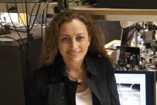
Adela Ben-Yakar
· ProfessorVerifiedUniversity of Texas at Austin · Electrical and Computer Engineering
Active 1993–2026
About
Dr. Adela Ben-Yakar is the Cockrell Family Chair in Engineering in the Department of Electrical and Computer Engineering, Department of Mechanical Engineering, and Department of Biomedical Engineering at The University of Texas at Austin. She received her Ph.D. from Stanford University in Engineering and completed postdoctoral work at Stanford and Harvard Universities in Physics. Her research focuses on the development of novel technologies for therapeutic and diagnostic applications of ultrafast lasers, the development of the fastest fluorescence microscopy systems, high-throughput microfluidic systems for high-content screening of model organisms and organoids, and the application of machine learning to biomedical optical microscopy. Dr. Ben-Yakar has published more than 70 peer-reviewed journal articles, many in high-impact journals such as Nature, Nature Methods, and Nature Communications, and holds 8 issued and 7 pending patents. She is a Fellow of SPIE, Optica, and AIMBE, and has received numerous awards including the Fulbright Scholarship, Zonta Amelia Earhart Award, NSF Career Award, Human Frontier Science Program Research Award, NIH Director’s Transformative Award, and Faculty Investment Initiative Program Fellowship. She has presented over 120 plenary and invited talks worldwide and is the co-founder and CEO of vivoVerse, a startup providing cost-effective and rapid testing of drugs and chemicals using a high-throughput microfluidic imaging platform integrated with AI-enabled data analytics.
Research signals
Five dimensions sourced from public faculty / publication signals. Sign in to compare against your own profile and see your match score.
Research topics
- Materials science
- Optics
- Physics
- Optoelectronics
- Chemistry
- Biomedical engineering
- Nanotechnology
- Medicine
Selected publications
Planar laser activated neuronal scanning (PLANS) for high-speed flow cytometry using a pencil beam
2026-03-05
articleSenior authorHigh-speed ultrafast laser surgery scalpel: design, validation, and evaluation
2026-03-04
articleSenior authorUltrafast laser ablation offers unparalleled spatial and thermal confinement, making it a compelling candidate for high-precision spinal bone surgery. However, the inherently low ablation rates of ultrafast lasers and the challenges associated with probe miniaturization have significantly impeded their clinical translation. In this work, we present the design and prototyping of a new-generation, fiber-delivered ultrafast laser surgical scalpel optimized for both minimally invasive and open spinal procedures. The system integrates high-efficiency laser delivery via a Kagome hollow-core photonic crystal fiber, a piezoelectric fiber-scanning mechanism for compact beam steering, a high-demagnification miniaturized objective for enhanced ablation efficiency, and an opto-mechanical architecture enabling controlled depth-wise material removal and handheld operation. Prior efforts in parametric optimization informed the selection of pulse duration, spot size, and field-of-view to enable ablation rates exceeding 20 mm<sup>3</sup>/min under high-average-power operation. The optical design achieves a focused spot radius of 7.87 μm with a working distance of 3.8 mm, suitable for deep spinal bone incisions. Preliminary handheld tests on bone and synthetic spine models demonstrate stable focal-plane ablation, ergonomic maneuverability, and access to confined anatomical regions. These results establish a scalable and clinically translatable platform for ultrafast laser–based spinal surgery and represent a significant step toward replacing conventional mechanical bone removal tools.
Overcoming thermal constraints in two-photon imaging using thermoelectric cooling
2026-03-04
articleSenior authorTwo-photon laser scanning microscopy is widely used for deep-tissue imaging but is increasingly constrained by bulk tissue heating arising from linear absorption of excitation light. This volumetric heating accumulates with imaging depth, imposing conservative safe power limits that restrict achievable signal-to-noise ratio and imaging performance. While passive mitigation strategies such as objective are commonly employed, their effectiveness in suppressing bulk heating is limited. Here, we present a coupled numerical and experimental framework to quantify laser-induced bulk tissue heating during two-photon imaging and to evaluate active thermoelectric cooling strategies under realistic imaging conditions. Optical energy deposition is modeled using Monte Carlo simulations of photon transport and coupled to transient bioheat transfer calculations solved using an implicit finite difference scheme. The model is formulated in a two-dimensional domain and incorporates generalized thermal boundary conditions to represent localized thermoelectric cooling. The framework is validated against contact thermometry measurements performed in tissue-mimicking phantoms during 1035 nm point-scanning excitation at imaging depths of 300 μm and 1000 μm. Lateral temperature profiling demonstrates strong agreement between simulated and measured temperature rise, with an average error below 8%. Using a peak temperature rise criterion of 2°C, depth-dependent safe average power thresholds are quantified in the absence of active cooling. Applying thermoelectric cooling under otherwise identical imaging conditions substantially relaxes these thermal constraints, enabling up to a 4× increase in deliverable average power at shallow imaging depths and a 3.67× increase at deeper imaging depths. These results demonstrate the potential of active thermoelectric cooling to mitigate bulk tissue heating and extend the operational envelope of high-power two-photon microscopy.
2026-03-05
articleTwo-Photon Line Excitation Array Detection (2p-LEAD) is a novel, high-speed imaging method designed to overcome the speed limitations of traditional multi-photon microscopy. While conventional point-scanning is restricted to sub-30 Hz frame rates, 2p-LEAD can achieve 4 kHz frame rates at a 250 μm x 96 μm field-of-view. By coupling galvanometric line scanning with a parallelized 32-channel photomultiplier tube (PMT) array, we have constructed one of the fastest twophoton microscopes. Our system maintains the critical balance of subcellular resolution, kHz temporal resolution, and signal-to-noise ratio (SNR) through several key features. Temporal focusing confines the point-spread function (PSF) axially to reduce out-of-focus background, while the 32-channel PMT array enables highly efficient, parallelized photon collection. Additionally, optimizations of the optical configuration, excitation conditions, and detection hardware drastically improve the SNR for a given laser power, thereby mitigating the risk of phototoxicity and photodamage during sensitive, long-duration <i>in vivo</i> experiments. We demonstrate this advancement in capability by imaging the mammalian brain <i>in vivo</i>, resolving highly dynamic neurovascular events. The system's combination of high spatial resolution, temporal resolution, and SNR enables the quantitative measurement of fast-flowing red blood cells through cortical capillaries without motion artifacts. This demonstrated performance establishes a robust platform for future upgrades that may help enable comprehensive 4D investigation of functional and hemodynamic dynamics throughout large volumes of the mouse brain.
2026-03-04
articleSenior author2026-03-04
articleOpen accessSenior authorUltrafast laser surgery presents a promising alternative to conventional surgical tools in procedures where high precision and thermal safety are paramount. However, clinical translation has been limited by low ablation speeds and challenges in system integration. In this work, we investigate the optimization of bone ablation performance using a compact ultrafast laser delivery benchtop system based on Kagome hollow‑core fiber and piezo‑enabled beam scanning. We systematically evaluate the influence of spot size, fluence, and pulse overlap on ablation efficiency. Our results reveal a strong dependence of threshold fluence on the number of overlapping pulses, with smaller spot sizes yielding higher ablation efficiencies. Ablation rates >20 mm<sup>3</sup>/min has been achieved with 1 ps laser pulse at 11.8 W, highlighting a clear pathway toward achieving the clinically required rate of 1 mm<sup>3</sup>/s. Additionally, we report the first successful submerged ultrafast ablation of hard tissue using picosecond pulses and present initial thermal‑profiling data to assess heat accumulation during high‑power high-repetition rate ablation. Together, these findings provide key design considerations for the development of clinically viable, miniaturized ultrafast laser bone‑surgery probes.
Inverse-scattering in biological samples via beam-propagation
bioRxiv (Cold Spring Harbor Laboratory) · 2025-08-22 · 2 citations
preprintOpen accessMultiple scattering limits optical imaging in thick biological samples by scrambling sample-specific information. Physics-based inverse-scattering methods aim to computationally recover this information, often using non-convex optimization to reconstruct the scatter-corrected sample. However, this non-convexity can lead to inaccurate reconstructions, especially in highly scattering samples. Here, we show that various implementation strategies for even the same inverse-scattering method significantly affect reconstruction quality. We demonstrate this using multi-slice beam propagation (MSBP), a relatively simple nonconvex inverse-scattering method that reconstructs a scattering sample's 3D refractive-index (RI). By systematically conducting MSBP-based inverse-scattering on both phantoms and biological samples, we showed that an amplitude-only cost function in the inverse-solver, combined with angular and defocus diversity in the scattering measurements, enabled high-quality, fully-volumetric RI imaging. This approach achieved subcellular resolution and label-free 3D contrast across diverse, multiple-scattering samples. These results lay the groundwork for robust use of inverse-scattering techniques to achieve biologically interpretable 3D imaging in increasingly thick, multicellular samples, introducing a new paradigm for deep-tissue computational imaging.
2025-03-20
articleSenior authorTo numerically characterize optothermal interactions at the laser-tissue interface during tissue imaging or surgery, the bioheat transfer partial differential equation (PDE) must be solved using a laser excitation source function. Given the range of possible experimental scenarios, the PDEs become parametric and must be solved for multiple optical and laser parameters. Due to constraints such as solver stability and domain size, classical numerical techniques used for performing such simulations are often computationally taxing and time consuming. To overcome these challenges, we propose to learn the mapping between infinite dimensional function spaces of the parameterized laser source functions and the solutions of the parametric bioheat transfer PDEs. In this work, we introduce Res-DeepONet, a new neural network architecture leveraging deep operator networks for rapidly accelerating PDE inferences. We train our operator network using synthetic laser heating source functions consisting of Gaussian beam profiles. The proposed neural network architecture demonstrates excellent generalization performance on unseen Gaussian source functions, achieving a 30× speedup over the classical finite difference method. With the introduction of Res-DeepONets, we aim to bring a paradigm shift in performing bioheat transfer simulations in the realm of laser-tissue interactions.
bioRxiv (Cold Spring Harbor Laboratory) · 2025-06-06 · 3 citations
preprintOpen accessSenior authorCorrespondingHigh-content imaging (HCI) and analysis are the keys for advancing our understanding of the science behind organogenesis. To this end, culturing adult stem cell-derived organoids (ASOs) in a platform that also enables live imaging, staining, immobilization, and fast high-resolution imaging is crucial. However, existing platforms only partially satisfy these requirements. In this study, we present the OrganoidChip+, an all-in-one microfluidic device designed to integrate both culturing and HCI of ASOs all within one platform. We previously developed the OrganoidChip as a robust imaging tool. Now, the OrganoidChip+ incorporates several additional features for culturing organoids in addition to fluorescence staining and imaging without the need for sample transfer. The organoids grown within a culture chamber are stained and then transferred to immobilization chambers for blur-free, high-resolution imaging at predetermined locations. We cultured adult stem cell-derived intestinal organoids in the chip for 7 days and tracked growth rates of each organoid using intermittent brightfield images, followed by multiple image-based assays, including viability assay using widefield fluorescence imaging, a redox ratio assay using label-free, two-color, two-photon microscopy, and immunofluorescence assays using confocal microscopy. These assays serve as proof-of-concept to showcase the chip's capabilities in HCI of ASOs. Organoids cultured in the chip exhibited superior average growth rates over those in traditional Matrigel dome cultures, off-chip. Viability and redox ratio measurements of on-chip organoids were comparable or slightly better than their off-chip counterparts. Confocal imaging further confirmed that the OrganoidChip+ supports robust organoid culture while enabling detailed, high-resolution analysis. This all-in-one platform holds great potential for advancing ASO-based research, offering a scalable and cost-effective solution for HCI and analysis in organogenesis, drug screening, and disease modeling.
Evaluating Seeding Density Effects on Cardiac Organoid Health and Functionality for Toxicity Studies
Tissue Engineering Part A · 2025-11-03 · 1 citations
articleOpen accessDevelopment of relevant human induced pluripotent stem cell-derived cardiac organoids is essential to recapitulate myocardium physiology and functionality for the assessment of drug-induced toxicity evaluations. However, the optimal conditions for culturing self-aggregating multicellular cardiac organoids are not well-elucidated, particularly the impact of noncardiomyocytes. In this study, we generated cardiac organoids at varying seeding densities to formulate organoids that meet or exceed the biological diffusion limit. We assessed their morphology, gene expression profiles, beating functionality, viability, and mitochondrial activity over time. Our results show that organoid sizes stabilize by 7 days of culture, regardless of seeding density. However, organoids seeded with 20,000 cells retained a more optimal cardiac signature that promotes cardiac maturity and minimizes fibrotic tendencies, especially when cultured for longer than 7 days. While all organoid populations maintained their beating functionalities, those seeded with 80,000 cells exhibited greater cell shedding and increased apoptosis at long-term culture. In contrast, minimal apoptosis was observed in organoids seeded with 20,000 cells after 7 days. Mitochondrial staining further revealed that organoids seeded with 20,000 cells consistently demonstrated higher metabolic activity. Taken together, organoids seeded with 20,000 cells and cultured for 7 days yielded the healthiest morphology, transcriptional signature, and viability while maintaining robust beating kinetics. Importantly, the organoid model identified in this study demonstrated a selectivity index (SI) that is over an order of magnitude larger than that of two-dimensional cultures, showing improved sensitivity to clinically relevant doxorubicin-induced cardiotoxicity, enabling more accurate dose-response evaluations that better reflect therapeutic conditions.
Recent grants
NIH · $1.7M · 2012
NIH · $152k · 2008
CAREER: Plasmonic Laser Nanosurgery Using Molecularly Targeted Gold Nanoparticles
NSF · $400k · 2009–2015
NIH · $2.8M · 2016
NSF · $513k · 2005–2008
Frequent coauthors
- 25 shared
Nicholas J. Durr
Johns Hopkins University
- 22 shared
Ronald K. Hanson
- 17 shared
Liam Andrus
- 17 shared
Murat Yıldırım
Cleveland Clinic Lerner College of Medicine
- 16 shared
Christopher L. Hoy
- 14 shared
Kaushik Subramanian
The University of Texas at Austin
- 14 shared
Tomasz Tkaczyk
Rice University
- 14 shared
M. Kamel
National Water Research Center
Labs
Awards & honors
- Fellow of SPIE
- Fellow of Optica
- Fellow of AIMBE
- Fulbright Scholarship
- Zonta Amelia Earhart Award
- Resume-aware match score
- Save to shortlist
- AI-drafted outreach
See your match with Adela Ben-Yakar
PhdFit ranks faculty by your research interests, methods, and publications — grounded in their actual work, not templates.
- Free to start
- No credit card
- 30-second signup