Resume-aware faculty matching

Find professors who actually fit you

Upload your resume. Four AI agents analyze your background, rank the faculty who fit, inspect their recent research, and help you draft outreach — grounded in their actual work, not templates.

Free to startNo credit cardCancel anytime
Top matches Balanced preset
Dr. Sarah Chen
Stanford · Interpretability · NLP
91
Dr. Marcus Holloway
MIT · Robotics · RL
84
Dr. Aisha Okonkwo
CMU · Fairness · HCI
82
Nova · Professor Researcher · re-ranking top 20…
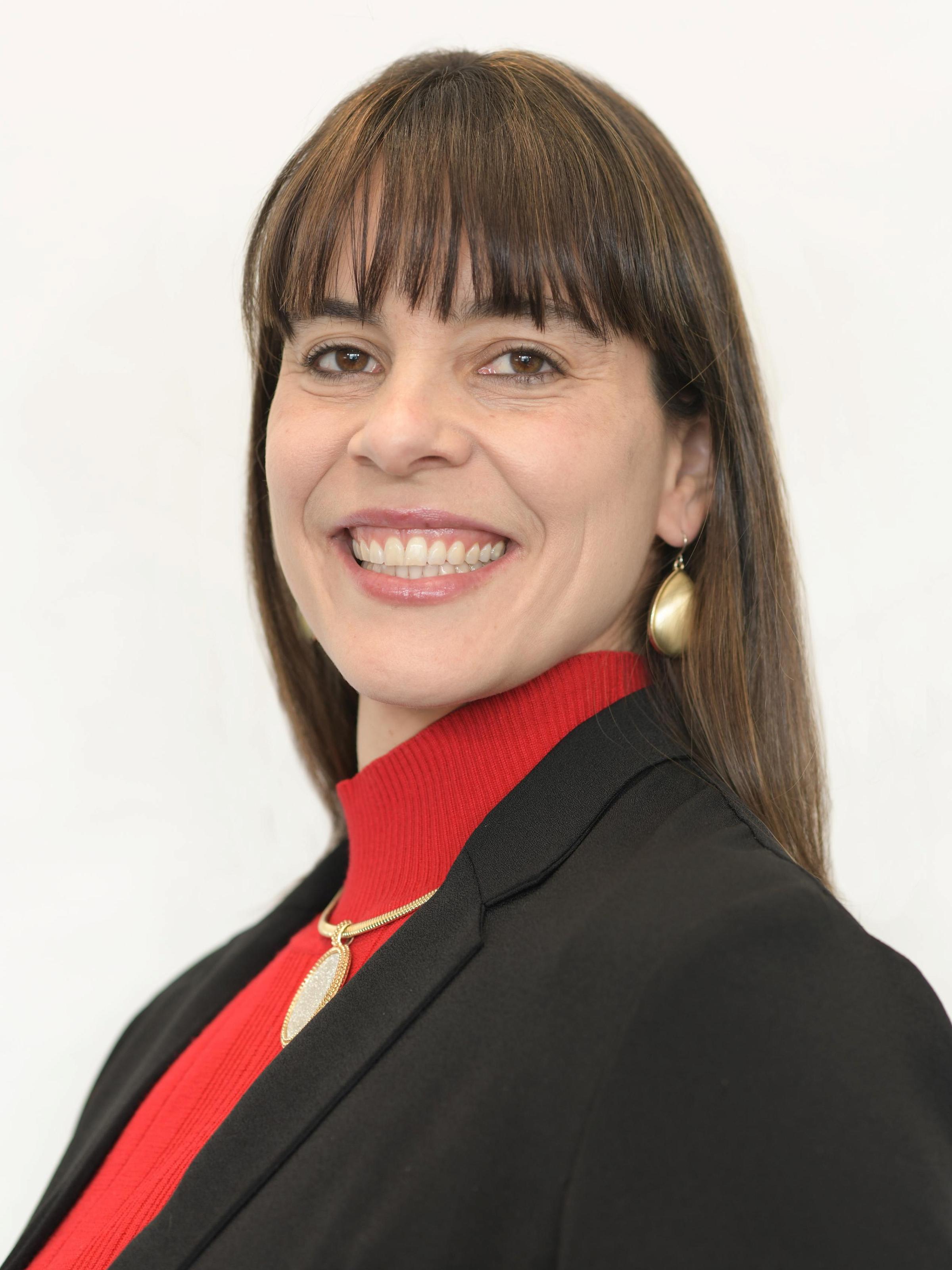
Ana Beatriz (Bia) DePaula-Silva

Ana Beatriz (Bia) DePaula-Silva

· Assistant Professor, Pharmacology and ToxicologyVerified

University of Utah · Department of Pharmacology & Toxicology

Active 2014–2026

h-index12
Citations572
Papers2914 last 5y
Funding
See your match with Ana Beatriz (Bia) DePaula-Silva — sign in to PhdFit.Sign in

About

Ana Beatriz (Bia) DePaula-Silva, PhD, is an Assistant Professor in the Department of Pharmacology & Toxicology at the College of Pharmacy. Her research focuses on how viral-host interactions are associated with the development of neuroinflammatory and neurodegenerative diseases, with particular emphasis on the involvement of CNS-infiltrating immune cells in conditions such as epilepsy and multiple sclerosis. Her work aims to shed light on disease mechanisms to facilitate the development of new therapies for neuroinflammatory and neurodegenerative conditions. DePaula-Silva's research includes studying the role of brain infiltrating immune cells, such as macrophages, in the development of temporal lobe epilepsy through a mouse model infected with Theiler’s Murine Encephalomyelitis virus, which leads to viral-induced epilepsy. Additionally, her work on multiple sclerosis involves understanding how viral-induced CD8 T cells contribute to axonal damage and demyelination using a mouse model of TMEV-induced demyelinating disease. Her academic background includes a BSc from Sao Paulo State University, a PhD in Microbiology and Immunology from the University of Utah, and postdoctoral training at the University of Utah. Her research contributes to understanding neuroinflammatory processes and aims to inform the development of therapeutic strategies.

Research topics

  • Biology
  • Immunology
  • Medicine
  • Neuroscience
  • Virology
  • Psychiatry
  • Intensive care medicine

Selected publications

  • The Ketogenic Diet Fails to Mitigate Seizures and Neuroinflammatory Responses in a Mouse Model of Virus-Induced Epilepsy

    bioRxiv (Cold Spring Harbor Laboratory) · 2026-05-21

    articleSenior authorCorresponding

    Objective: The ketogenic diet (KD) is a high-fat, low-carbohydrate intervention widely used to treat drug-resistant epilepsy, thought to reduce seizures through a combination of metabolic, neuronal, and microbiota-dependent mechanisms. Additionally, recent studies suggest that the anticonvulsant effects of KD require the gut microbiota, with taxa such as Akkermansia and Parabacteroides contributing to seizure protection by modulating host neurotransmitter balance and neural excitability. While KD has been shown to be effective in reducing seizure burden across different epilepsies, its antiseizure effect on infection-driven seizures, which are often driven by acute neuroinflammation, has not been evaluated. Here, we evaluated the effects of KD on seizure burden, neuroimmune responses, and gut microbiota composition in the Theiler's murine encephalomyelitis virus (TMEV) model of virus-induced epilepsy. Methods: Mice were maintained on either a KD or a normal diet prior to intracerebral TMEV infection. Seizures were induced by handling and scored twice daily from day 3 to 7 post-infection. Neuroimmune responses were assessed by flow cytometry, and fecal microbial composition was analyzed using 16S rRNA gene sequencing. Results: Despite achieving ketosis, KD did not reduce seizure incidence, seizure burden, or seizure severity during acute TMEV infection. KD also did not significantly alter overall immune cell infiltration into the central nervous system, indicating limited effects on global neuroinflammation. However, KD significantly reshaped the gut microbiota, reducing alpha diversity (richness, Shannon diversity, and evenness) and strongly altering community structure with clear separation between diet groups, including enrichment of taxa such as Akkermansia , Acetatifactor , Dorea ,and Flintibacter , and depletion of fiber-associated taxa including Bifidobacterium and Roseburia . However, these microbial shifts were insufficient to mitigate inflammation-driven seizures. Significance: These results demonstrate that KD's anticonvulsant efficacy is highly context-dependent, and that KD-driven changes in microbiota- and metabolite-mediated mechanisms may be ineffective against infection-associated epilepsy, suggesting that inflammation-driven seizures require distinct therapeutic approaches.

  • Investigating the Role of Cortical Microglia in a Mouse Model of Viral Infection-Induced Seizures

    eNeuro · 2026-02-01

    articleOpen access

    Microglia, resident immune sentinels in the brain, are crucial in responding to tissue damage, infection, damage signals like purines (ATP/ADP), and clearing cellular debris. It is currently unknown how microglial reactivity progresses and contributes to seizure development following Theiler's murine encephalomyelitis virus (TMEV) infection. Previously, it has been demonstrated that purinergic signaling in microglia is disrupted in the hippocampus of TMEV-infected mice. However, whether reactive cortical microglia also exhibit changes in purinergic signaling, cytokine levels, and purinergic receptors is unknown. Thus, we seek to evaluate region-based differences in microglial reactivity in the TMEV model. We employed a custom triple transgenic mouse line expressing tdTomato and GCaMP6f under a CX3CR1 Cre promoter and exogenously applied ATP/ADP to acute brain slice preparations from TMEV-infected mice and controls of either sex. Interestingly and in contrast to what is observed in the hippocampus, we found that despite microglial reactivity in the cortex, microglia can respond to purinergic damage signals and engage calcium signaling pathways, comparable to PBS controls. Using a cytokine panel, we also found that proinflammatory cytokine levels (TNF-α, IL-1α, and IFN-γ) are brain region dependent in mice infected with TMEV. Using RNAscope FISH, we observed increases in expression of purinergic receptors responsible for microglial motility (P2Y 12 R) and inflammation (P2X 7 R) in the cortex. Collectively our results suggest that following TMEV infection, microglial response to novel damage signals, as well as the production of proinflammatory cytokines, varies as a function of the brain region.

  • Viral encephalitis and seizures cause rapid depletion of neuronal progenitor cells and alter neurogenesis in the adult mouse dentate gyrus

    Frontiers in Cellular Neuroscience · 2025-01-14 · 2 citations

    articleOpen access

    Infections impacting the central nervous system (CNS) constitute a substantial predisposing factor for the emergence of epileptic seizures. Given that epilepsy conventionally correlates with hippocampal sclerosis and neuronal degeneration, a potentially innovative avenue for therapeutic intervention involves fostering adult neurogenesis, a process primarily occurring within the subgranular zone of the dentate gyrus (DG) through the differentiation of neural stem cells (NSC). While experimental seizures induced by chemoconvulsants or electrical stimulation transiently enhance neurogenesis, the effects of encephalitis and the resultant virus-induced seizures remain inadequately understood. Thus, this study employed the Theiler's Murine Encephalomyelitis Virus (TMEV) model of virus-induced seizures in adult C57BL/6J mice to investigate the impact of infection-induced seizures on neurogenesis at three distinct time points [3, 7, and 14 days post-infection (dpi)]. Immunohistochemical analysis revealed a reduction in the overall number of proliferating cells post-infection. More notably, the specific cell types exhibiting proliferation diverged between TMEV and control (CTR) mice: (1) Neuronal progenitors (doublecortin, DCX + ) were almost entirely absent at 3 dpi in the dorsal DG. They resumed proliferation at 14 dpi, but, did not recover to CTR levels, and displayed aberrant migration patterns. (2) The number of proliferating NSCs significantly decreased within the dorsal DG of TMEV mice at 14 dpi compared to CTR, while (3) a heightened population of proliferating astrocytes was observed. Most observed changes were not different between seizing and non-seizing infected mice. In summary, our findings demonstrate that viral infection rapidly depletes neuronal progenitor cells and causes aberrant migration of the remaining ones, potentially contributing to hyperexcitability. Additionally, the increased differentiation toward glial cell fates in infected mice emerges as a possible additional pro-epileptogenic mechanism.

  • Investigating the role of cortical microglia in a mouse model of viral infection-induced seizures

    bioRxiv (Cold Spring Harbor Laboratory) · 2025-08-27

    preprintOpen access

    Microglia, resident immune sentinels in the brain, are crucial in responding to tissue damage, infection, damage signals like purines (ATP/ ADP), and clearing cellular debris. It is currently unknown how microglial reactivity progresses and contributes to seizure development following Theilers Murine Encephalomyelitis Virus (TMEV) infection. Previously, our group has demonstrated that purinergic signaling in microglia is disrupted in the hippocampus of TMEV-infected mice. However, whether reactive cortical microglia also exhibit changes in purinergic signaling, cytokine levels, and purinergic receptors are unknown. Thus, we seek to evaluate region-based differences in microglial reactivity in the TMEV model. We employed a custom triple transgenic mouse line expressing tdTomato and GCaMP6f under a CX3CR1 Cre promoter and exogenously applied ATP/ADP to acute brain slice preparations from TMEV-infected mice and controls. Interestingly and in contrast to what is observed in hippocampus, we found that despite microglial reactivity in the cortex, microglia can respond to purinergic damage signals and engage calcium signaling pathways, comparable to PBS controls. Using a cytokine panel, we also found that pro-inflammatory cytokine levels (TNF-α, IL-1α and IFN-γ) are brain-region dependent in mice infected with TMEV. Using RNAScope-FISH, we observed increases in expression of purinergic receptors responsible for microglial motility (P2Y12R) and inflammation (P2X7R) in the cortex. Collectively our results suggest that following TMEV infection, microglial response to novel damage signals, as well as the production of proinflammatory cytokines, varies as a function of brain region.

  • Brain on Fire: How Brain Infection and Neuroinflammation Drive Worldwide Epilepsy Burden

    Epiliepsy currents/Epilepsy currents · 2024-04-30 · 3 citations

    reviewOpen access

    Roughly 80% of the global burden of epilepsy resides in low- and middle-income countries (LMICs; WHO, 2022). Despite numerous new therapies for the treatment of epilepsy, the number of patients who remain resistant to available medications is unchanged. Additionally, no therapy has yet been clinically proven to prevent or attenuate the development of epilepsy in at-risk individuals. Unfortunately, access to next generation therapies in LMICs is low, the stigma associated with epilepsy remains high, and access to adequate resources is unchanged. Thus, the global epilepsy burden disproportionately falls on LMICs such that strategies to conscientiously integrate global epilepsy risk factors into preclinical research may meaningfully advance 21st century epilepsy therapies. Brain infections are one of the main risk factors for epilepsy in resource-poor settings. Further, both infection- and autoimmune-associated encephalitis contribute to worldwide epilepsy risk and remain relatively understudied. For example, clinical SARS CoV-2 infection can induce rare instances of encephalopathy and acute seizures. Among viruses known to cause acute brain infection, enteroviruses increase risk for encephalitis-induced epilepsy, but are not associated with risk for other neurodevelopmental disorders (eg, autism spectrum or attentional deficit hyperactivity disorders). Naturally occurring models of viral infection-induced epilepsy therefore provide an exquisite opportunity to uncover novel contributors to epileptogenesis. Moreover, the convergent neuroinflammatory pathways that are associated with viral infection-induced encephalitis and autoimmune encephalitis reflect an untapped therapeutic opportunity to meaningfully reduce the global burden of epilepsy. This review summarizes the latest advances in translational research integrating encephalitis-induced seizure and epilepsy models, in tandem with progress in clinical diagnosis of inflammation and virally mediated epilepsy. This improved awareness of the shared biological underpinnings of epileptogenesis following brain infection or autoimmune encephalitis is anticipated to beneficially impact the global burden of epilepsy.

  • The Contribution of Microglia and Brain-Infiltrating Macrophages to the Pathogenesis of Neuroinflammatory and Neurodegenerative Diseases during TMEV Infection of the Central Nervous System

    Viruses · 2024-01-13 · 22 citations

    articleOpen access1st authorCorresponding

    The infection of the central nervous system (CNS) with neurotropic viruses induces neuroinflammation and is associated with the development of neuroinflammatory and neurodegenerative diseases, including multiple sclerosis and epilepsy. The activation of the innate and adaptive immune response, including microglial, macrophages, and T and B cells, while required for efficient viral control within the CNS, is also associated with neuropathology. Under healthy conditions, resident microglia play a pivotal role in maintaining CNS homeostasis. However, during pathological events, such as CNS viral infection, microglia become reactive, and immune cells from the periphery infiltrate into the brain, disrupting CNS homeostasis and contributing to disease development. Theiler's murine encephalomyelitis virus (TMEV), a neurotropic picornavirus, is used in two distinct mouse models: TMEV-induced demyelination disease (TMEV-IDD) and TMEV-induced seizures, representing mouse models of multiple sclerosis and epilepsy, respectively. These murine models have contributed substantially to our understanding of the pathophysiology of MS and seizures/epilepsy following viral infection, serving as critical tools for identifying pharmacological targetable pathways to modulate disease development. This review aims to discuss the host-pathogen interaction during a neurotropic picornavirus infection and to shed light on our current understanding of the multifaceted roles played by microglia and macrophages in the context of these two complexes viral-induced disease.

  • The CSF1R-Microglia Axis Has Protective Host-Specific Roles During Neurotropic Picornavirus Infection

    Frontiers in Immunology · 2021 · 20 citations

    • Biology
    • Immunology
    • Virology

    Viral encephalitis is a major cause of morbidity and mortality, but the manifestation of disease varies greatly between individuals even in response to the same virus. Microglia are professional antigen presenting cells that reside in the central nervous system (CNS) parenchyma that are poised to respond to viral insults. However, the role of microglia in initiating and coordinating the antiviral response is not completely understood. Utilizing Theiler's murine encephalomyelitis virus (TMEV), a neurotropic picornavirus, and PLX5622, a small molecule inhibitor of colony-stimulating factor 1 receptor (CSF1R) signaling that can deplete microglia in the CNS; we investigated the role of the CSF1R-microglia axis in neurotropic picornavirus infection of C57BL/6J and SJL/J mice. These mouse strains differ in their ability to clear TMEV and exhibit different neurological disease in response to TMEV infection. CSF1R antagonism in C57BL/6J mice, which normally clear TMEV in the CNS, led to acute fatal encephalitis. In contrast, CSF1R antagonism in SJL/J mice, which normally develop a chronic CNS TMEV infection, did not result in acute encephalitis, but exacerbated TMEV-induced demyelination. Immunologically, inhibition of CSF1R in C57BL/6J mice reduced major histocompatibility complex II expression in microglia, decreased the proportion of regulatory T cells in the CNS, and upregulated proinflammatory pathways in CNS T cells. Acute CSF1R inhibition in SJL/J mice had no effect on microglial MHC-II expression and upregulated anti-inflammatory pathways in CNS T cells, however chronic CSF1R inhibition resulted in broad immunosuppression. Our results demonstrate strain-specific effects of the CSF1R-microglia axis in the context of neurotropic viral infection as well as inherent differences in microglial antigen presentation and subsequent T cell crosstalk that contribute to susceptibility to neurotropic picornavirus infection.

  • Figure 3C datasource file.xlsx

    Figshare · 2021-01-01

    datasetOpen access

    phospho CDK1(Y15) protein levels in 293FT cells the presence or absence of Vpr

  • Figure 4A .fcs datasource files

    Figshare · 2021-01-01

    datasetOpen access

    Figure 4A. BV421 fluorescence from HeLa cells treated with DMSO or 1-10 microM CDK1 inhibitor Ro3306.

  • Figure 3B .fcs filesource data

    Figshare · 2021-01-01

    datasetOpen access

    Figure 3B. BV421 fluorescence data from HeLa cells treated with either scramble, CCDC137 or MCM10 siRNA.

Frequent coauthors

  • Robert S. Fujinami

    20 shared
  • Jane E. Libbey

    University of Utah

    19 shared
  • Vicente Planelles

    University of Utah

    19 shared
  • Daniel J. Doty

    University of Utah

    15 shared
  • John Michael S. Sanchez

    Cedars-Sinai Smidt Heart Institute

    13 shared
  • Laura Martins

    University of Utah

    13 shared
  • Tyler J. Hanak

    NuVasive (United States)

    8 shared
  • Alberto Bosque

    George Washington University

    5 shared

Education

  • Postdoctoral fellow, Pathology

    University of Utah

  • Ph.D., Pathology

    University of Utah

    2015
  • B.Sc. in Pharmacy and Biochemistry

    Sao Paulo State University

    2008
  • Resume-aware match score
  • Save to shortlist
  • AI-drafted outreach

See your match with Ana Beatriz (Bia) DePaula-Silva

PhdFit ranks faculty by your research interests, methods, and publications — grounded in their actual work, not templates.

  • Free to start
  • No credit card
  • 30-second signup