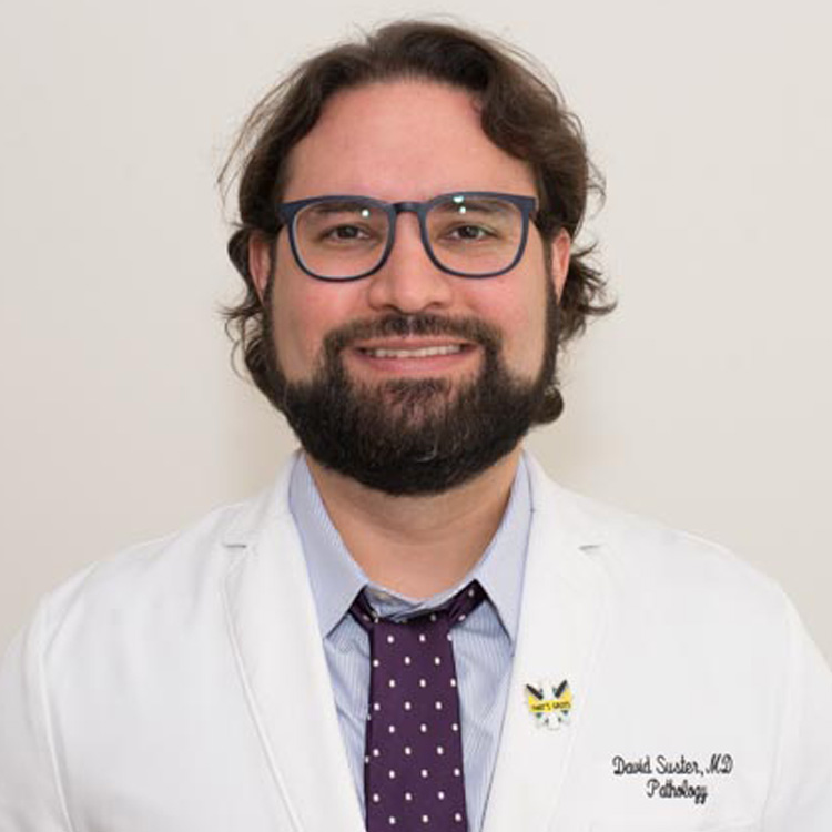
David Suster
· Associate ProfessorVerifiedRutgers University · Pathology, Immunology and Laboratory Medicine
Active 2005–2026
About
Dr. David I. Suster is an Associate Professor in the Department of Pathology, Immunology and Laboratory Medicine at Rutgers New Jersey Medical School. He earned his M.D. from Tulane University School of Medicine in 2014 and completed his pathology residency at Beth Israel Deaconess Medical Center, Harvard Medical School. Following his residency, he completed a fellowship at Massachusetts General Hospital, Harvard Medical School, specializing in bone and soft tissue pathology as well as pulmonary pathology. His areas of interest include bone and soft tissue pathology and pulmonary pathology. Dr. Suster has contributed to the field through numerous publications, including research on cartilaginous lesions of bone, sarcomatoid renal cell carcinoma, osteoblastoma, spindle cell liposarcoma, and thymic carcinomas, among others. His work involves detailed diagnostic and research efforts in these areas, reflecting his expertise in complex pathological diagnoses and research in soft tissue and pulmonary diseases.
Research topics
- Medicine
- Pathology
- Biology
- Biochemistry
- Genetics
- Molecular biology
- Endocrinology
- Virology
- Immunology
- Cell biology
Selected publications
80 Osteosarcoma in Adults Over 40: A Clinicopathologic Study of 58 Patients
Laboratory Investigation · 2026-03-01
article1st authorCorrespondingLaboratory Investigation · 2026-03-01
articleOpen accessSenior authorLaboratory Investigation · 2026-03-01
article1st authorCorrespondingChondroblastoma with prominent secondary aneurysmal bone cyst-like changes: The role of H3.3 K36 M
Human Pathology · 2026-04-27
article1st authorCorrespondingLaboratory Investigation · 2026-03-01
articleSenior authorJournal of Clinical Pathology · 2025-08-22
article1st authorCorrespondingAIMS: ) in a large number of soft tissue neoplasms using a tissue microarray technique. METHODS: 489 cases of soft tissue neoplasms, including benign and malignant entities, were collected from the files of the respective institutions and constructed into tissue microarrays. Tissue microarrays were stained for ERG immunohistochemistry using two antibodies, EP111 and EPR3864. RESULTS: A total of 25 cases (5.1%) were identified that were positive for ERG using the monoclonal antibody EP111 and 15 cases (3%) using the monoclonal antibody EPR3864, including rhabdomyosarcoma, peripheral nerve sheath tumours, synovial sarcoma, myxofibrosarcoma, epithelioid sarcoma, dermatofibrosarcoma protuberans, low-grade fibromyxoid sarcoma, nodular fasciitis and dedifferentiated liposarcoma. The most consistently stained tumours included synovial sarcoma, rhabdomyosarcoma and benign and malignant peripheral nerve sheath tumours. Various other fibroblastic proliferations, including dermatofibrosarcoma protuberans, myxofibrosarcoma, low-grade fibromyxoid sarcoma and nodular fasciitis, also showed positive staining in a small fraction of cases. One case of dedifferentiated liposarcoma showed nuclear positivity for ERG, and one case of epithelioid sarcoma was also positive. CONCLUSIONS: This study supports the value of ERG as a highly sensitive and specific marker for the diagnosis of vascular neoplasms but also demonstrates rare cases of aberrant staining and underscores the need to assess soft tissue tumours using a panel of stains and interpret the results of immunohistochemistry in the appropriate histological and clinical context.
Pseudoneoplastic Fat Atrophy of Intra-Abdominal Sites
The American Journal of Surgical Pathology · 2025-12-17
article1st authorCorrespondingPatients undergoing periods of starvation or malnutrition may develop degenerative and/or atrophic changes of various organs. These findings may manifest histologically and may cause confusion when first encountered, raising a differential that includes benign and malignant tumor entities. Herein, we report 16 cases of incidentally identified degenerative changes within abdominal-site fatty tissues that caused diagnostic difficulties. The patients were 11 males and 5 females, aged from 1 day to 82 years (mean: 58 years). The degenerative changes were all incidentally identified within adipose tissue of the abdominal cavity or adjacent to organs contained within the abdominal cavity in procedures done for unrelated reasons such as cancer, infection, or perforation. Cases were identified in the colon (n=6), small intestine (n=4), omentum (n=3), inguinal hernia sac (n=1), peritoneum (n=1), and retroperitoneal soft tissue (n=1). Patients had variable past medical histories with multiple comorbidities. Ten patients presented with some form of malnutrition and/or cachexia. Histologically, the lesions demonstrated lobules of atrophic-appearing fat with small signet-ring-like adipocytes that were sometimes bilobed. The nodules varied in size and were characterized in some cases by a variably myxoid stromal background, often with a prominent delicate capillary network. Immunohistochemistry was performed and was uniformly positive for S100 protein and negative for cytokeratins. The lesions were generally recognized as degenerative, but various differential diagnoses proposed at the time of sign-out included signet-ring cell carcinoma, liposarcoma, and vascular lesions, among others. Nomenclature to describe this phenomenon has been inconsistent in the literature, and thus, we suggest the term "pseudoneoplastic fat atrophy."
49 Expanding the Clinicopathologic Spectrum of Sarcomas with EWSR1::SSX Fusions
Laboratory Investigation · 2025-03-01 · 1 citations
articleOpen accessOssifying Spindled and Epithelioid Tumor: A Novel Soft Tissue Tumor
Modern Pathology · 2025-07-16 · 5 citations
articleJournal of Molecular Diagnostics · 2025-05-15
review
Frequent coauthors
- 88 shared
Saul Suster
Medical College of Wisconsin
- 42 shared
Alexander C. Mackinnon
University of Alabama at Birmingham
- 21 shared
Gerard J. Nuovo
The Ohio State University Wexner Medical Center
- 20 shared
Germán Pihán
- 20 shared
John Gross
Johns Hopkins Medicine
- 19 shared
Natali Ronen
Medical College of Wisconsin
- 18 shared
Esmerina Tili
The Ohio State University Wexner Medical Center
- 18 shared
Haider A. Mejbel
Emory University
Education
- 2014
M.D.
Tulane University School of Medicine
Other, bone and soft tissue and pulmonary pathology
Massachusetts General Hospital, Harvard Medical School
Other, pathology
Beth Israel Deaconess Medical Center, Harvard Medical School
- Resume-aware match score
- Save to shortlist
- AI-drafted outreach
See your match with David Suster
PhdFit ranks faculty by your research interests, methods, and publications — grounded in their actual work, not templates.
- Free to start
- No credit card
- 30-second signup