Resume-aware faculty matching

Find professors who actually fit you

Upload your resume. Four AI agents analyze your background, rank the faculty who fit, inspect their recent research, and help you draft outreach — grounded in their actual work, not templates.

Free to startNo credit cardCancel anytime
Top matches Balanced preset
Dr. Sarah Chen
Stanford · Interpretability · NLP
91
Dr. Marcus Holloway
MIT · Robotics · RL
84
Dr. Aisha Okonkwo
CMU · Fairness · HCI
82
Nova · Professor Researcher · re-ranking top 20…
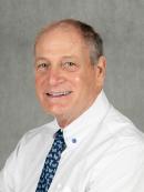
Howard Drew

Howard Drew

· Professor

Rutgers University · Periodontics

Active 1987–2023

h-index8
Citations160
Papers191 last 5y
Funding
See your match with Howard Drew — sign in to PhdFit.Sign in

Research topics

  • Medicine
  • Dermatology
  • Pathology

Selected publications

  • Novel treatment approach of localized juvenile spongiotic gingival hyperplasia with use of Nd:YAG laser

    Clinical Advances in Periodontics · 2023 · 4 citations

    Senior authorCorresponding
    • Medicine
    • Pathology
    • Dermatology

    INTRODUCTION: This article describes the management of two patients who presented with localized juvenile spongiotic gingival hyperplasia. This disease entity is poorly understood, and literature regarding successful treatment is scant. However, there are common themes to management including proper diagnosis and treatment via removal of the affected tissue. The biopsy demonstrates intercellular edema and neutrophil infiltrate with epithelial and connective tissue disease components, so surgical deepithelialization may not be adequate to definitively treat the disease. METHODS AND RESULTS: This article presents two cases of the disease and suggests the use of the Nd:YAG laser as an alternative management modality. CONCLUSION: To our knowledge, we present the first cases of localized juvenile spongiotic gingival hyperplasia treated with the Nd:YAG laser. KEY POINTS: Why are these cases new information? To our knowledge, this case series illustrates the first use of an Nd:YAG laser to treat the rare pathology localized juvenile spongiotic gingival hyperplasia. What are the keys to successful management of these cases? To properly manage this rare presentation, proper diagnosis is paramount. Following diagnosis with microscopic evaluation, deepithelialization and treatment of underlying connective tissue infiltrate via the Nd:YAG laser offers an elegant option to treat the pathology and maintain esthetic outcomes. What are the primary limitations to success in these cases? The primary limitations of these cases include the small sample size, which is attributable to the rarity of the disease.

  • Novel Approach to Overcome Limitations Associated With Immediate Implant Placement: Osteotome Technique.

    PubMed · 2019-10-01

    article

    This case series introduces a novel osteotome technique as an alternative to conventional drilling protocols for immediate fixtures. Immediate implant placement is associated with significantly less bone-to-implant contact, and navigating the anatomy of the socket can be difficult. Osteotomy drills remove already-limited bone following atraumatic extractions. The osteotome technique has been introduced as an alternative approach to prepare the osteotomy in the appropriate position relative to the socket apex and surrounding alveolar walls. The surgical technique and case reports demonstrating it are presented, highlighting the technique's benefits.

  • Lip Repositioning with Vestibular Shallowing Technique for Treatment of Excessive Gingival Display with Various Etiologies

    The International Journal of Periodontics & Restorative Dentistry · 2018-01-01 · 12 citations

    article

    Alireza Torabi, DDS, MS/Babak Najafi, DDS, MDS, FRCD(C)/Howard J. Drew, DMD/Emil G. Cappetta, DMD: Improvement of smile esthetics is a major goal of modern dentistry. Various treatment modalities have been proposed to correct excessive gingival display (EGD), depending on the identified etiologies. This study reports on the clinical and patient-centered outcomes of a novel lip repositioning technique with vestibular shallowing approach in the treatment of three types of EGD with varying etiologies. Periosteal fenestration with cicatrization (scarification) was performed at the mucogingival junction to ensure the stability of esthetic outcomes. Suspensory triangular sutures and extraoral tissue stabilization tapes were used to facilitate the cicatrization process during the healing phase of this novel technique. Postoperative clinical examination revealed 84% reduction in gingival display that remained stable for 13 to 16 months with a high level of patient satisfaction.

  • Amniotic Band Syndrome: A Multidisciplinary Care Approach to the Treatment of a Rare Case

    The Cleft Palate-Craniofacial Journal · 2018-04-06 · 4 citations

    article

    Amniotic band syndrome (ABS) is a rare developmental disorder associated with defects such as syndactyly, facial and/or palatal clefts, and dental anomalies like malformed or impacted teeth. In this report, a patient with ABS was successfully treated with orthodontic, endodontic, and periodontal therapies. Cone beam computed tomography revealed a unique eruptive path of the impacted central incisor through the incisive canal and cleft area. The tooth was occlusally and functionally stable after 1 year of treatment. Multidisciplinary care was critical to the success of this case. Available literature on ABS is also briefly reviewed.

  • Surgical Treatment of Advanced Peri‐Implantitis With a Dual Augmentation Technique: A Report of Two Cases With 15‐Month Follow‐Up

    Clinical Advances in Periodontics · 2016-06-20 · 2 citations

    article

    Introduction: With the prevalence of peri‐implantitis increasing, management of the disease is an important therapy to consider. The goal of a dual augmentation technique in the treatment of peri‐implantitis is to address hard and soft tissue deficiency simultaneously. This report of two cases presents clinical and radiographic outcomes for surgical treatment of peri‐implantitis with a dual augmentation technique in advanced peri‐implantitis cases with inadequate keratinized tissue. Case Series: Two patients diagnosed with advanced peri‐implantitis exhibiting bleeding on probing (BOP), probing depth (PD) >6 mm, and bone loss >3 mm were treated. Affected implants were treated with specific surface decontamination protocol followed by using a combination of recombinant human platelet‐derived growth factor with a mixture of freeze‐dried bone allograft and deproteinized bovine‐derived bone. Subsequently, acellular dermal matrix was used as a membrane to cover defects and augment soft tissue volume at the same time. The dual augmentation technique demonstrated significant radiographic bone gain in 3 to 5 months and increase in mucosal thickness that appeared to be stable during the follow‐up period of 15 months. Surgical reentry provided further evidence to confirm radiographic bone fill. Clinical examination also revealed significant reduction in PD and BOP. Conclusion: Within the limitations of this report, treatment of peri‐implantitis with a dual augmentation technique appeared to enhance bone and soft tissue regeneration in advanced peri‐implantitis cases with existing mucogingival defects.

  • Comparison of Osteotome and Conventional Drilling Techniques for Primary Implant Stability: An In Vitro Study

    Journal of Oral Implantology · 2016-03-03 · 28 citations

    article

    It may be difficult to achieve primary stability in the posterior maxilla because of poor quality and quantity of bone. Studies have shown that the osteotome technique immediately increases bone density thereby increasing primary stability. An in vitro study was conducted to compare the stability achieved by the osteotome and conventional drilling techniques in low density bone. Forty endosseous implant fixtures (n = 40) were inserted in a solid rigid polyurethane block simulating low density (D3) bone. The implants were divided into 4 groups to test 2 variables: (1) implant length (10 mm or 13 mm) and (2) preparation of osteotomy (conventional drilling or osteotome technique). Insertion torque (IT) and resonance frequency analysis (RFA) were measured for each implant. Statistical analysis using one-way ANOVA and Tukey post hoc test was done to study IT and RFA data of the 4 groups. Pearson Correlation test was used to determine the correlation between IT and RFA values of the implants. The IT and RFA values were statistically significant higher using the osteotome technique as compared to conventional drilling (P < 0.0001). Statistically significant higher values were also found for IT and RFA of 13 mm implants as compared to 10 mm implants. A significant correlation was found between insertion torque and RFA values in all 4 groups (r = 0.86, P < 0.0001). The conclusion was that the osteotome technique significantly increased primary stability.

  • Space Maintenance Using Tenting Screws in Atrophic Extraction Sockets

    Journal of Oral Implantology · 2016-02-29 · 4 citations

    articleSenior author

    The alveolar process is a tooth dependent tissue that develops with tooth eruption. The volume of the alveolar process is dependent on the shape, size, inclination of the tooth, and its eruption pattern. The tooth is anchored by bundle bone, which is invaded by periodontal fibers.1Following tooth extraction, the bundle bone is rapidly resorbed resulting in loss of height and width.2 The loss of width of alveolar bone is the major concern and is approximately 3 times greater than the loss of height.4,5 The buccal plate resorbs more because it is thinner, averaging about 0.8mm in the anterior teeth region and 1.1mm at the premolar sites.6Approximately 50% of the alveolar ridge width is lost after tooth extraction, most of which occurs during the first 3 months.7The rationale for socket grafting is to have bone available to place implants in proper position for a successful implant prosthesis8 and studies have stated that the survival rate of implants placed in grafted bone is similar to those in native bone.9Extraction sockets with deficient buccal bone that are classified as Funato's Class III and IV types,10 or Elian's Type III11 are difficult to predictably graft.Treatment options include guided bone regeneration (GBR) vs titanium reinforced membrane, a titanium mesh, or a high-density polytetrafluoroethylene (PTFE) membrane, or block auto/allogenous grafts.The tenting screw technique (TST) is an alternative to these procedures and is as effective in promoting new bone formation, localized to the site that is highly predictable, time efficient, cost effective, with less patient morbidity.TST is based on the basic principles of GBR utilizing resorbable barrier membranes. This technique provides and maintains space, allowing stabilization of the blood clot during healing.This article illustrates 3 clinical case reports utilizing the tenting screw technique to facilitate GBR in atrophic extraction sockets.A standard surgical protocol was followed. Patients were locally anesthetized using 2% lidocaine with 1:100 000 epinephrine. A crestal incision was made slightly toward the lingual/palatal in an attempt to preserve keratinized gingiva and limit secondary intention healing over the site. A full-thickness flap was elevated. The tooth was extracted using periotomes. Sockets were degranulated using curettes.A tenting screw osteotomy was created with the tenting screw kit drill. The tenting screw was placed vertically, horizontally, or diagonally depending on where augmentation was needed.Salvin bone fixation screw kit was used, which contains 2 latch type or straight handpiece drills of different sizes, self-tapping titanium screws, and a locking tip screw driver. Titanium tenting screws are available in different sizes of 4mm, 6mm, 8mm, 10mm, 12mm, and 15mm in length and 1.5mm to 2mm in diameter. If primary stability is difficult, a longer screw can be used. The lingual/buccal plate can be perforated to get bicortical stabilization.The vertical height of the screw is determined by proximal height of the adjacent bone. The screw head can be placed 1–2mm above this height if required (Figure 1a and b). Single or multiple screws can be used based on the size of defect.The buccal vestibular periosteum is scored, and, if necessary, horizontal cut back incisions are made to achieve tension-free primary closure. A membrane template is fabricated prior to membrane placement, extending at least 3mm onto the sound buccal and palatal/lingual bone.The extraction socket is decorticated to create the necessary heme for osteogenesis. Particulate allograft condensed into the socket and followed by placement of the membrane. Membrane-tacking screws are recommended only with multiple-site socket preservation or in conjunction with ridge augmentation. Tension-free primary closure is desired whenever possible.Postoperatively, all patients were placed on antibiotics, chlorhexidine rinse, and antiinflammatories. A methylprednisolone dose pack was given only with multiple site augmentation or ridge augmentation.After 3 months sites were reentered for placement of the implant. Tenting screws were removed by gently unscrewing in a counterclockwise direction. Implant placement was then carried out in the usual manner.With GBR, the mucogingival junction shifts coronally in an attempt to achieve primary closure. Hence, we apically positioned the flap during uncovery to eliminate the need for additional soft tissue grafting.A 51-year-old male, with nonsignificant medical history, presented with pain and mobility of his right central incisor. Clinical and radiographic exam revealed a hopeless right central incisor (#8), with a hemi-septal defect on the mesial and a compromised buccal plate of bone (Figure 2a). Although teeth #7 and #9 were treatment planned for full coverage restorations, the patient preferred 3 individual restorations rather than a fixed partial denture option. The patient was treatment planned for extraction of tooth #8, with tenting screw GBR.After flap reflection and extraction of the tooth, significant loss of facial and mesial bone were noted (Figure 2b). After degranulation of the defect, the tenting screw was placed in the center of the alveolus, angled buccally at the level of the adjacent interproximal bone heights. (Figures 2c and d). Decortication was done to obtain the necessary bleeding for angiogenesis followed by condensation of cancellous allograft (Maxxeus Dental, Kettering, Ohio) of particle size 0.25–1mm (Figure 2e). Resorbable bilayer collagen membrane (Geistlich Bio-Gide, Princeton, NJ) was placed buccal to palatal. Tension-free primary closure was obtained, and the area was fixed provisional to prevent premature loading of the graft site (Figure 2f). At 4 months, a follow-up radiograph was taken, showing radiographic bone fill to the level of the screw head (Figure 2g). Adequate bone formation for implant placement occurs as early as 12 weeks following tooth extraction, with insignificant changes in alveolar ridge dimensions.12A 67-year-old male with a noncontributory medical history presented for comprehensive dental care. The mandibular right first molar was deemed hopeless due to an endo-perio lesion (Figure 3a). The area was treatment planned for implant crowns in areas #29 and #30. Upon reflection and extraction of the tooth, it was found that both the buccal and interradicular bone was compromised. The tenting screw was placed buccally in the defect (Figure 3b). Particulate cancellous bone graft (Maxxeus) of size 0.25–1mm and resorbable collagen membrane (Geistlich Bio-Gide) were placed and tension-free primary closure was achieved (Figures 3c–e). At 4 months, 2 platform-switched 3i/Biomet fixtures were placed (Figure 3f). The final restorations were placed with sufficient embrasure spacing as well as an adequate band of attached keratinized tissue (Figure 3g).A 41-year-old healthy female presented with a chief complaint of wanting “implants to replace her missing teeth.” Tooth #4 revealed an endodontically hopeless tooth. The tooth was treatment planned for tenting screw GBR and implant placement. Upon flap reflection and extraction of the tooth, significant loss of vertical bone was noted, and a tenting screw was placed vertically in the center of the defect with the screw slightly coronal to the adjacent interproximal bone heights. Cancellous bone allograft (Maxxeus) of particle size 0.25–1mm was used to fill the socket and regenerate the missing buccal plate. Resorbable collagen membrane (Geistlich Bio-Gide) was trimmed to the desired size and placed from buccal to palatal.Figure 4a–d shows the radiographic sequence of the preop radiograph, tenting screw placement, immediate grafting with the screw, and 4 months postoperative healing after tenting screw GBR.Figure 4e shows the radiographic progression of implant placement at the time of surgery, parallel pin, test body implant, and placement of a tapered implant (4/3mm × 11.5mm Biomet 3i implant). The implant was placed at crest in the ridge area (Figure 4f and g). Although no histology was done, clinical impression was of Type III bone. Figure 4h and i shows good crestal bone level at 3 years postoperatively.Implant surgery is a restoratively driven procedure. Atrophic extraction sockets with defects need to be grafted in order to have a successful esthetic, functional, and restorative outcome. A common problem encountered in the regeneration of these defects is membrane collapse resulting in the loss of graft volume. Various surgical procedures including autogenous/allogenic block grafts and titanium mesh with or without growth factors have been used successfully in the past.The advantage of the tenting screw technique is its space-making ability. During the healing period, the diagonally placed tenting screws provide a tenting effect and resist the collapse of the membrane, thus maintaining the volume and geometry of the space. This allows for the stabilization of the blood clot, which is fundamental for success.Some advantages of the tenting screw technique include the following:The overall success of this procedure depends on proper design and reflection of tissue flaps, meticulous degranulation, and tension-free primary closure. Although many clinicians advocate flap less extractions, large defects with severe buccal bone discrepancy need to be flapped and thoroughly degranulated for successful regeneration.Tension-free primary closure is obtained by scoring of the periosteum, which also promotes angiogenesis by causing bleeding into the graft material.13Extraction results in significant horizontal and vertical bone resorption. The horizontal bone loss is much higher than the vertical bone loss. The resorption of the buccal plate is more than that of the lingual plate due to a more rapid resorption rate during the first 3–6 months after tooth extraction.Various socket preservation techniques can be successfully employed to minimize the extent of bone resorption, although bone loss cannot be prevented completely.GBR using tenting screws is an alternative method to the gold standard of block grafting. It can be used successfully in the regeneration of atrophic extraction sockets.It is a highly predictable, cost-effective procedure with less healing time and patient morbidity. It should be considered as one of the treatment options in the management of atrophic extraction socket.

  • Novel Techniques with the Aid of a Staged CBCT Guided Surgical Protocol

    Case Reports in Dentistry · 2015-01-01 · 13 citations

    articleOpen accessSenior author

    The case report will present some novel techniques for using a "staged" protocol utilizing strategic periodontally involved teeth as transitional abutments in combination with CBCT guided implant surgery. Staging the case prevented premature loading of the grafted sites during the healing phase. A CBCT following a tenting screw guided bone regeneration procedure ensured adequate bone to place an implant fixture. Proper assessment of the CBCT allowed the surgeon to do an osteotome internal sinus lift in an optimum location. The depth of the bone needed for the osteotome sinus floor elevation was planned. The staged appliance allowed these sinus-augmented sites to heal for an extended period of time compared to implants, which were uncovered and loaded at an earlier time frame. The staged protocol and CBCT analysis enabled the immediate implants to be placed in proper alignment to the adjacent fixture. After teeth were extracted, the osseointegrated implants were converted to abutments for the transitional appliance. Finally, the staged protocol allowed for soft tissue enhancement in the implant and pontic areas prior to final insertion of the prosthesis.

  • The Significance of Attached Gingiva Around Teeth and Implants.

    PubMed · 2015-01-01 · 1 citations

    article
  • Reconstruction of the Narrow Ridge Using Combined Ridge Split and Guided Bone Regeneration with rhPDGF-BB Growth Factor–Enhanced Allograft

    The International Journal of Periodontics & Restorative Dentistry · 2014-01-01 · 10 citations

    article

    In clinical situations where the presence of severe horizontal ridge deficiencies precludes simultaneous implant placement and bone augmentation, a staged approach may be desirable to allow optimal implant placement. Numerous therapeutic options are available for the treatment of the horizontally deficient ridge. With advances in tissue engineering, the use of growth factors can significantly improve wound healing with more rapid bone formation and maturation. These case reports demonstrate a technique that enhances the predictability of horizontal bone gain with reduced surgical trauma and postoperative complications. Recombinant human platelet-derived growth factor BB (rhPDGF-BB) in combination with particulate allograft is used to stimulate the proliferation and migration of osteogenic cells. A ridge split technique with vertical bone incisions allows expansion and mobilization of the buccal plate, creating a space that will contain the particulate graft material. Decortication of the mobilized buccal plate will create pathways to allow cellular and vascular access for enhanced maturation. Additional graft material is placed lateral to the mobilized buccal plate to increase apical ridge width. The use of piezoelectric surgery enables a precise crestal bony incision in severely deficient ridge widths and aids in faster wound healing. This study discusses the technique and the recommended therapeutic considerations to ensure predictable regeneration of adequate bone for optimal implant placement in horizontally deficient ridges.

Frequent coauthors

  • Emil Cappetta

    4 shared
  • B Simon

    3 shared
  • Babak Najafi

    3 shared
  • Tat Fai Chiang

    3 shared
  • Oscar Frank

    2 shared
  • Sofia D Petrov

    2 shared
  • Walter Molofsky

    2 shared
  • Herman Baker

    Rutgers, The State University of New Jersey

    2 shared

Labs

  • PeriodonticsPI

  • Resume-aware match score
  • Save to shortlist
  • AI-drafted outreach

See your match with Howard Drew

PhdFit ranks faculty by your research interests, methods, and publications — grounded in their actual work, not templates.

  • Free to start
  • No credit card
  • 30-second signup