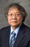
Hua Hua Chang
· Professor of Statistics (Courtesy), Charles R. Hicks Chair Person, College of EducationVerifiedPurdue University · Statistics
Active 1991–2025
About
Hua Hua Chang is a Professor of Statistics (Courtesy) and holds the Charles R. Hicks Chair Person in the College of Education at Purdue University. His professional contact information includes an email address at chang606@purdue.edu and an office located in BRNG 5166. Further details about his academic profile can be found on his faculty webpage at Purdue University. The biography provided does not include specific information about his research focus, background, or key contributions.
Research topics
- Biology
- Cell biology
- Biophysics
- Genetics
- Endocrinology
- Internal medicine
- Biochemistry
- Geometry
Selected publications
Drosophila photoreceptor tethering by a laminin-Eys scaffold
iScience · 2025-05-22 · 1 citations
articleOpen accessSenior author), a photoreceptor-secreted proteoglycan essential for IRS formation, guides LanB1 and perlecan deposition by cone cells during pupal development. Disruption of LanB1 results in rhabdomere tip detachment, IRS collapse, and impaired tension transmission. These findings reveal that cone cells and photoreceptors collaboratively sculpt a rigid LanB1 grid that caps and reinforces the distal IRS lumen. This composite ECM structure preserves rhabdomere organization and evenly distributes mechanical forces, ensuring photoreceptor alignment and optical fidelity.
Interommatidial cells build a tensile collagen network during Drosophila retinal morphogenesis
Current Biology · 2023-05-19 · 8 citations
articleOpen accessSenior authorAuxilin regulates intestinal stem cell proliferation through EGFR
Stem Cell Reports · 2022-04-14 · 8 citations
articleOpen accessAdult tissue homeostasis is maintained by residential stem cells. The proliferation and differentiation of adult stem cells must be tightly balanced to avoid excessive proliferation or premature differentiation. However, how stem cell proliferation is properly controlled remains elusive. Here, we find that auxilin (Aux) restricts intestinal stem cell (ISC) proliferation mainly through EGFR signaling. aux depletion leads to excessive ISC proliferation and midgut homeostasis disruption, which is unlikely caused by defective Notch signaling. Aux is expressed in multiple types of intestinal cells. Interestingly, aux depletion causes a dramatic increase in EGFR signaling, with a strong accumulation of EGFR at the plasma membrane and an increased expression of EGFR ligands in response to tissue stress. Furthermore, Aux co-localizes and associates with EGFR. Finally, blocking EGFR signaling completely suppresses the defects caused by aux depletion. Together, these data demonstrate that Aux mainly safeguards EGFR activation to keep a proper ISC proliferation rate to maintain midgut homeostasis.
De novo endocytic clathrin coats develop curvature at early stages of their formation
Developmental Cell · 2021 · 52 citations
- Biology
- Cell biology
- Biophysics
Development · 2021 · 21 citations
Senior authorCorresponding- Biology
- Cell biology
- Biophysics
Actomyosin contraction shapes the Drosophila eye's panoramic view. The convex curvature of the retinal epithelium, organized in ∼800 close-packed ommatidia, depends upon a fourfold condensation of the retinal floor mediated by contraction of actin stress fibers in the endfeet of interommatidial cells (IOCs). How these tensile forces are coordinated is not known. Here, we discover a previously unobserved phenomenon: Ca2+ waves regularly propagate across the IOC network in pupal and adult eyes. Genetic evidence demonstrates that IOC waves are independent of phototransduction, but require the inositol 1,4,5-triphosphate receptor (IP3R), suggesting that these waves are mediated by Ca2+ releases from endoplasmic reticulum stores. Removal of IP3R disrupts stress fibers in IOC endfeet and increases the basal retinal surface by ∼40%, linking IOC waves to facilitation of stress fiber contraction and floor morphogenesis. Furthermore, IP3R loss disrupts the organization of a collagen IV network underneath the IOC endfeet, implicating the extracellular matrix and its interaction with stress fibers in eye morphogenesis. We propose that coordinated cytosolic Ca2+ increases in IOC waves promote stress fiber contractions, ensuring an organized application of the planar tensile forces that condense the retinal floor. This article has an associated 'The people behind the papers' interview.
The people behind the papers – Donald Ready and Henry Chang
Development · 2021-11-15
articleOpen access1st authorCorrespondingABSTRACT Coordinating contractility across tissues is key for maintaining the fidelity of morphogenetic processes. A new paper in Development explains how cytosolic calcium waves in the interommatidial cells, the pigment-secreting cells in the Drosophila eye, lead to remodelling of the retinal floor, by activating contraction of the basal actomyosin stress fibres. We caught up with the authors, Professor Donald Ready and Associate Professor Henry Chang, both from Purdue University, to find out more about this story.
Nature Communications · 2021-07-12 · 12 citations
articleOpen accessAbstract The maintenance of constant karyoplasmic ratios suggests that nuclear size has physiological significance. Nuclear size anomalies have been linked to malignant transformation, although the mechanism remains unclear. By expressing dominant-negative TER94 mutants in Drosophila photoreceptors, here we show disruption of VCP (valosin-containing protein, human TER94 ortholog), a ubiquitin-dependent segregase, causes progressive nuclear size increase. Loss of VCP function leads to accumulations of MDC1 (mediator of DNA damage checkpoint protein 1), connecting DNA damage or associated responses to enlarged nuclei. TER94 can interact with MDC1 and decreases MDC1 levels, suggesting that MDC1 is a VCP substrate. Our evidence indicates that MDC1 accumulation stabilizes p53A, leading to TER94 K2A -associated nuclear size increase. Together with a previous report that p53A disrupts autophagic flux, we propose that the stabilization of p53A in TER94 K2A -expressing cells likely hinders the removal of nuclear content, resulting in aberrant nuclear size increase.
bioRxiv (Cold Spring Harbor Laboratory) · 2021-04-09 · 1 citations
preprintOpen accessSenior authorCorrespondingAbstract Actomyosin contraction shapes the Drosophila eye’s panoramic view. The convex curvature of the retinal epithelium, organized in ∼800 close-packed ommatidia, depends upon a fourfold condensation of the retinal floor mediated by contraction of actin stress fibers in the endfeet of inter o mmatidial c ells (IOCs). How these tensile forces are coordinated is not known. Here, we discover a novel phenomenon: Ca 2+ waves regularly propagate across the IOC network in pupal and adult eyes. Genetic evidence demonstrates that IOC Ca 2+ waves are independent of phototransduction, but require inositol 1,4,5-triphosphate receptor (IP3R), suggesting these waves are mediated by Ca 2+ releases from ER stores. Removal of IP3R disrupts stress fibers in IOC endfeet and increases the basal retinal surface by ∼40%, linking IOC waves to facilitating stress fiber contraction and floor morphogenesis. Further, IP3R loss disrupts the organization of a collagen IV network underneath the IOC endfeet, implicating ECM and its interaction with stress fibers in eye morphogenesis. We propose that coordinated Ca 2+ spikes in IOC waves promote stress fiber contractions, ensuring an organized application of the planar tensile forces that condense the retinal floor. Summary Statement Ca 2+ waves have an important role in generating tensile forces to shape the Drosophila eye’s convex curvature. Coordinated Ca 2+ spikes facilitate actomyosin contractions at the basal endfeet of interommatidial cells.
Nature Communications · 2020 · 76 citations
- Cell biology
- Biology
- Genetics
Mitochondrial aging, which results in mitochondrial dysfunction, is strongly linked to many age-related diseases. Aging is associated with mitochondrial enlargement and transport of cytosolic proteins into mitochondria. The underlying homeostatic mechanisms that regulate mitochondrial morphology and function, and their breakdown during aging, remain unclear. Here, we identify a mitochondrial protein trafficking pathway in Drosophila melanogaster involving the mitochondria-associated protein Dosmit. Dosmit induces mitochondrial enlargement and the formation of double-membraned vesicles containing cytosolic protein within mitochondria. The rate of vesicle formation increases with age. Vesicles originate from the outer mitochondrial membrane as observed by tracking Tom20 localization, and the process is mediated by the mitochondria-associated Rab32 protein. Dosmit expression level is closely linked to the rate of ubiquitinated protein aggregation, which are themselves associated with age-related diseases. The mitochondrial protein trafficking route mediated by Dosmit offers a promising target for future age-related mitochondrial disease therapies.
UNC Libraries · 2020-11-10
articleOpen accessThe cell surface proteoglycan, syndecan-1, is essential for normal epithelial morphology and function. Syndecan-1 is selectively localized to the basolateral domain of polarized epithelial cells and interacts with cytosolic PDZ (PSD-95, discs large, ZO-1) domain-containing proteins. Here, we show that the polarity of syndecan-1 is determined by its type II PDZ-binding motif. Mutations within the PDZ-binding motif lead to the mislocalization of syndecan-1 to the apical surface. In contrast to previous examples, however, PDZ-binding motif-dependent polarity is not determined by retention at the basolateral surface but rather by polarized sorting prior to syndecan-1’s arrival at the plasma membrane. Although none of the four known PDZ-binding partners of syndecan-1 appears to control basolateral localization, our results show that the PDZ-binding motif of syndecan-1 is decoded along the biosynthetic pathway establishing a potential role for PDZ-mediated interactions in polarized sorting.
Frequent coauthors
- 22 shared
Gerald M. Rubin
Howard Hughes Medical Institute
- 19 shared
Felix Karim
Amgen (United States)
- 15 shared
Marc Therrien
Institute for Research in Immunology and Cancer
- 14 shared
David A. Wassarman
University of Wisconsin–Madison
- 13 shared
Noah M. Solomon
- 9 shared
Ira Mellman
- 8 shared
Tanya Wolff
Columbia University
- 7 shared
E. Malavasi
Education
- 1995
PhD, Molecular & Cell Biology
University of California Berkeley
- 1991
BA, Biochemical Sciences
Harvard College
- Resume-aware match score
- Save to shortlist
- AI-drafted outreach
See your match with Hua Hua Chang
PhdFit ranks faculty by your research interests, methods, and publications — grounded in their actual work, not templates.
- Free to start
- No credit card
- 30-second signup