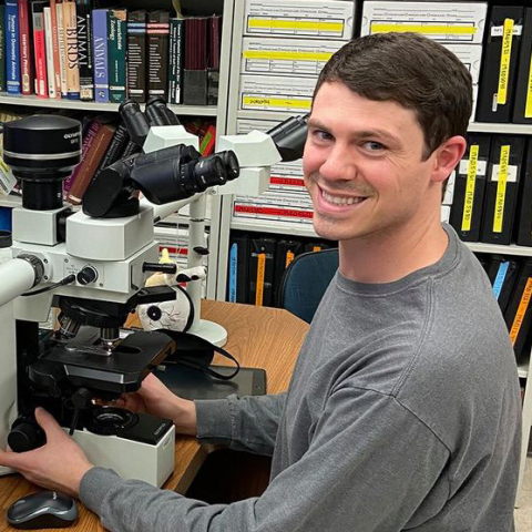
Ryan Taylor
· Assistant Clinical ProfessorCornell University · Public Health and Food Safety
Active 1975–2026
About
Ryan Taylor, DVM, DACVP, is an Assistant Clinical Professor in the Department of Population Medicine and Diagnostic Sciences at Cornell University College of Veterinary Medicine in Ithaca, NY. He earned his Doctor of Veterinary Medicine degree from Mississippi State University in 2017 and completed a veterinary anatomic pathology residency at Mississippi State University from 2017 to 2020. He is a Diplomate of the American College of Veterinary Pathology, having achieved this designation in 2021, and completed an ocular pathology fellowship at the Comparative Ocular Pathology Laboratory of Wisconsin (COPLOW) from 2020 to 2022. His research and clinical interests focus on ocular pathology. Ryan Taylor is actively involved in professional organizations such as the American College of Veterinary Pathology and the Society for Comparative Ocular Pathology (SCŌP). He serves as the primary editor and organizer of the Veterinary Ocular Pathology Atlas (VOPA). His contributions to veterinary pathology include numerous publications on ocular and other veterinary pathological conditions, emphasizing his expertise in this specialty.
Research topics
- Medicine
- Biology
- Anatomy
- Obstetrics
- Pathology
- Surgery
- Dermatology
Selected publications
Veterinary Pathology · 2026-05-12
articleCarcinomas have been reported to metastasize to the globe, specifically the uvea; however, the distinguishing histologic features of intraocular metastatic carcinomas in cats are not well-documented. Eleven cases of feline ocular biopsies with intraocular metastatic carcinomas and history of a pulmonary mass submitted to the Comparative Ocular Pathology Laboratory of Wisconsin from 2019 to 2025 were collected and reviewed. Alcian blue stains and immunohistochemical markers for thyroid transcription factor-1 and vimentin were evaluated. No significant sex or breed predispositions were identified. Uveitis, glaucoma, and the presence of an ocular mass were the most common clinical reasons for enucleation. All cases had a thoracic mass identified by radiographs prior to enucleation. All neoplasms had carpeting to gland-forming growth patterns lining surfaces of intraocular structures, affecting the anterior uvea. Goblet cells were highlighted with Alcian blue staining in 7/11 cases (64%). Neoplastic cells immunolabeled for thyroid transcription factor-1 in 2/11 cases (18%) (neither of which had goblet cells) and vimentin in 4/11 cases (36%). Based on this study, feline intraocular metastatic neoplasms are commonly associated with glaucoma and less commonly with uveitis. Histomorphologic and immunohistochemical features of pulmonary origin neoplasia were not consistently found in these metastatic masses of suspected pulmonary origin; the presence of goblet cells was the most commonly identified histologic feature.
Journal of Veterinary Cardiology · 2026-02-02
articleVeterinary Ophthalmology · 2025-09-04
articleOBJECTIVE: To describe the clinical and histologic features of a sarcoid arising from the medial caruncle with spread to the third eyelid of a horse. ANIMAL STUDIED: A 20-year-old Quarter Horse gelding. PROCEDURES: An incisional biopsy of the medial caruncle mass was initially performed, with histopathology suggesting a sarcoid or other spindle cell neoplasm. The mass grew rapidly following the biopsy, expanding to affect the third eyelid and adjacent superior palpebral conjunctiva. A complete third eyelid and medial caruncle excision with resection of affected palpebral conjunctiva and adjunctive cryotherapy was subsequently performed. RESULTS: Histopathology revealed the mass to be consistent with an equine sarcoid as evidenced by a densely cellular population of neoplastic spindle cells arranged into interlacing bundles with collagenous stroma and with hyperplastic overlying epithelium extending into the neoplasm as thin rete ridges. Over 90% of neoplastic cells showed strong hybridization signaling for bovine papillomavirus types 1 and 2, supporting an underlying viral cause consistent with the diagnosis of a sarcoid. CONCLUSIONS: This paper describes the first reported case of an equine sarcoid arising from the medial caruncle with spread to the third eyelid, treated with third eyelid and medial caruncle excision and adjunctive cryotherapy. Veterinarians evaluating horses with masses affecting the medial caruncle and third eyelid should consider sarcoids as a differential diagnosis. Rapid growth of cutaneous sarcoids following incisional biopsy is common, and the same may be true for sarcoids that arise from the medial caruncle or third eyelid.
Pyovagina and sepsis due to <i>Streptococcus didelphis</i> in a Virginia opossum
Journal of Veterinary Diagnostic Investigation · 2025-02-12
articleOpen accessVirginia opossums ( Didelphis virginiana ) are the only native marsupials in the United States of America and Canada. Females have a reproductive tract consisting of a complex duplex (bifurcated) reproductive system, with 2 ovaries, 2 uteri and uterine tubes, and a vaginal complex composed of 2 lateral vaginae, vaginal sinuses, and a urogenital sinus. Few conditions of the reproductive tract have been described in these marsupials. A 2-y-old, female Virginia opossum was presented because of hematuria and was found dead shortly after admission. The autopsy revealed left lateral vaginal torsion and pyovagina. Streptococcus didelphis was isolated and identified from the lateral vaginal exudate, blood, and liver using matrix-assisted laser desorption/ionization time-of-flight mass spectrometry (MALDI-TOF MS). Histologic examination confirmed suppurative and lymphoplasmacytic vaginitis, endometritis, cystitis, pyelonephritis and interstitial nephritis, portal hepatitis with fibrosis, myocarditis, and adrenalitis with intralesional gram-positive cocci. These results suggest ascending S. didelphis infection of the lateral vagina with subsequent torsion, bacterial translocation, bacteremia, and sepsis as the cause of death.
Veterinary Record Case Reports · 2024-10-25 · 1 citations
articleOpen accessSenior authorAbstract A 13‐year‐old, domestic shorthair cat presented for evaluation of left‐sided exophthalmos, epiphora and third eyelid elevation of approximately 4‐month duration. Based on ophthalmic exam findings, a retrobulbar mass was suspected. Computed tomography of the head revealed a fluid to soft‐tissue, non‐contrast‐enhancing mass caudoventral to the left globe. Based on these findings, a retrobulbar abscess was considered the most likely differential, while a neoplastic mass could not be completely excluded. The mass and left globe were surgically excised via a modified lateral orbitotomy and exenteration. Histopathology and special staining of the mass was consistent with an orbital primitive neuroectodermal tumour, while the globe was unremarkable.
Optic nerve meningioma and cloacal adenocarcinoma in a Humboldt penguin
Journal of Veterinary Diagnostic Investigation · 2024-01-11
articleOpen access) was euthanized following a 3.5-mo history of weakened elimination mechanics, recurrent tenesmus, intermittent hemorrhagic droppings, and a cloacal mass. Blepharospasm, of unknown cause, of the right eye was present for ~3 mo before euthanasia. Autopsy revealed a cloacal adenocarcinoma with localized coelomic carcinomatosis and distant metastases to the liver and lungs. On histopathology, a 2.6 × 1.2 × 0.5-mm, well-demarcated mass was found surrounding the right optic nerve, expanding the subdural space and wrapping the leptomeninges. The mass was composed of neoplastic spindle-to-polygonal cells consistent with a meningioma, meningothelial subtype. No evidence of neoplasia was found in the optic chiasm or brain, indicating a primary retrobulbar meningioma. Immunohistochemistry for cytokeratin AE1/AE3, vimentin, and S100 revealed robust and consistent immunoreactivity to vimentin, and weak and variable immunoreactivity to cytokeratin and S100, supporting the diagnosis. Meningiomas have been described only rarely in avian species, and we found no reports of optic nerve meningiomas in any avian species to date. The optic nerve meningioma in this case was considered a clinically incidental finding.
2024-03-30
peer-reviewCraniofacial malformations in a stillborn mixed‐breed dog as a cause for dystocia
Veterinary Medicine and Science · 2024 · 1 citations
- Medicine
- Obstetrics
- Anatomy
This case report describes the findings of craniofacial anomalies associated with dystocia in a mixed-breed bitch. A bitch in labour was presented for evaluation of dystocia, and an emergency caesarean section was performed. Two pups with craniofacial abnormalities were delivered by hysterotomy. One pup was stillborn, with congenital anomalies including palatoschisis (cleft palate), cheiloschisis (cleft lip), an open fontanelle, and a narrow teardrop-shaped skull. The second pup was delivered alive and had cheiloschisis. Craniofacial malformations are a reported cause of dystocia in the dog, usually due to obstruction. However, dystocia in the reported case is presumed to have developed because the pup's craniofacial malformations prevented stimulation of uterine contractions. To the authors' knowledge, this is the first case report to describe craniofacial abnormalities affecting 100% of the litter and is the first known report to describe the relationship between craniofacial abnormalities and presumptive primary uterine inertia.
Journal of Exotic Pet Medicine · 2024-07-02
article2024-04-24
peer-review
Frequent coauthors
- 15 shared
Michael A. O. Lewis
Cardiff University
- 15 shared
Peter E. Bradley
National Institute of Standards and Technology
- 15 shared
Ray Radebaugh
Cryogenic Industries (United States)
- 12 shared
Gregory Nellis
University of Wisconsin–Madison
- 11 shared
S.A. Klein
Fraunhofer Research Institution for Energy Infrastructures and Geothermal Systems
- 5 shared
J. G. Weisend
- 4 shared
Mason Jager
Cornell University
- 4 shared
Callum G. Donnelly
University of California, Davis
Awards & honors
- CL Davis Veterinary Pathology Scholarship Award (2019)
- Class award for excellence in pathology (2017)
- Diplomate of American College of Veterinary Pathology (2021)
- Resume-aware match score
- Save to shortlist
- AI-drafted outreach
See your match with Ryan Taylor
PhdFit ranks faculty by your research interests, methods, and publications — grounded in their actual work, not templates.
- Free to start
- No credit card
- 30-second signup