Resume-aware faculty matching

Find professors who actually fit you

Upload your resume. Four AI agents analyze your background, rank the faculty who fit, inspect their recent research, and help you draft outreach — grounded in their actual work, not templates.

Free to startNo credit cardCancel anytime
Top matches Balanced preset
Dr. Sarah Chen
Stanford · Interpretability · NLP
91
Dr. Marcus Holloway
MIT · Robotics · RL
84
Dr. Aisha Okonkwo
CMU · Fairness · HCI
82
Nova · Professor Researcher · re-ranking top 20…
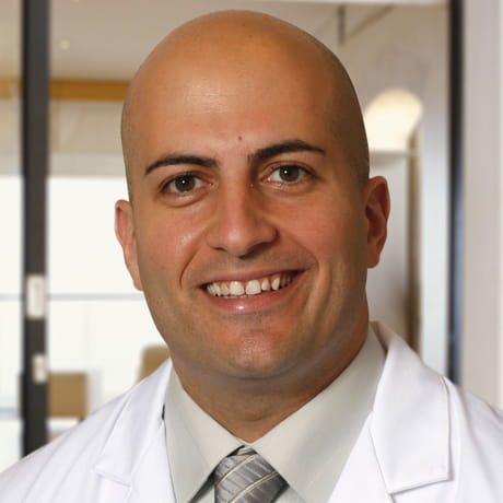
Said Atway

Said Atway

· Clinical Associate Professor of Orthopaedics, Division of Foot & Ankle Surgery, Section of PodiatryVerified

Ohio State University · Orthopaedics

Active 2008–2026

h-index8
Citations364
Papers3621 last 5y
Funding
See your match with Said Atway — sign in to PhdFit.Sign in

About

Said Atway, DPM, is a board-certified podiatric surgeon at The Ohio State University Wexner Medical Center, specializing in the diagnosis and treatment of a broad spectrum of foot and ankle conditions. His clinical expertise includes foot deformities such as bunions, flatfoot, and complex structural abnormalities, as well as plantar fasciitis, hallux valgus, and chronic wounds. He has advanced training in minimally invasive surgical techniques and innovative treatment approaches aimed at faster recovery, reduced postoperative pain, and improved long-term outcomes. His practice focuses on deformity correction, limb salvage, wound healing, and preventive strategies, with an emphasis on proactive treatment for patients of all ages, including young athletes and individuals seeking to maintain active lifestyles. In addition to his clinical work, Dr. Atway serves as an associate clinical professor in the department of orthopaedics at The Ohio State University College of Medicine and is the residency director for the podiatric medicine and surgery residency program. He is recognized for high patient satisfaction, being rated in the top 10 percent of physicians nationally in 2014, 2015, and 2016.

Research topics

  • Medicine
  • Surgery
  • Internal medicine
  • Nuclear medicine
  • Radiology
  • Orthodontics
  • Nursing
  • Endocrinology
  • Cardiology
  • Intensive care medicine

Selected publications

  • Abstract Th0077: Vessel-Specific CT Calcium Scoring in Peripheral Artery Disease Using Deep Learning

    JVS Vascular Science · 2026-01-01

    articleOpen access
  • Abstract Th0077: Vessel-Specific CT Calcium Scoring in Peripheral Artery Disease Using Deep Learning

    Arteriosclerosis Thrombosis and Vascular Biology · 2025-04-01

    article

    Objective: CT calcium scoring is a tool for assessing disease severity and risk for adverse events in coronary artery disease; however, quantification of vessel-specific calcium burden from CT images in peripheral artery disease (PAD) has remained relatively understudied due to the time-consuming nature of segmenting the arterial network. Therefore, we sought to test the performance of a semi-automated deep learning approach to segment and quantify vessel-specific calcium burden from CT images in PAD patients to streamline eventual clinical implementation of calcium scoring in PAD. Methods: Patients with PAD (N=80) were prospectively enrolled for non-contrast CT imaging. Images were manually segmented to quantify calcium mass for the femoral-popliteal, peroneal, anterior tibial, and posterior tibial arteries. Manually processed images were used as input data to train an nnU-Net deep learning model. Data augmentation techniques were applied to increase the dataset to 157 images (80 patients=157 legs) to achieve better generalization of results. The dataset was randomly split using an 90/10 ratio for model training and testing. Dice coefficient was calculated to assess the agreement between manual and deep learning image analysis results. Results: Deep learning-guided image segmentation results qualitatively agreed with manual image analysis ( Fig. 1A ). Quantitatively, deep learning produced dice coefficients of 0.82 ± 0.05 for femoral-popliteal, 0.71 ± 0.12 for anterior tibial, 0.74 ± 0.12 for posterior tibial, and 0.73 ± 0.12 for the peroneal artery, thus representing good performance for multi-vessel segmentation ( Fig. 1B ). Calcium mass values derived from both manual and deep learning image analysis demonstrated excellent agreement, with an intraclass correlation coefficient of 0.98 ( Fig. 1C ). Conclusions: Deep learning allows for accurate quantification of vessel-specific CT calcium values for the lower extremities of PAD patients, which is a challenging task due to arteries comprising a small percentage (i.e., 0.07%) of the overall CT image. This AI-based approach significantly reduces CT image analysis time from hours to seconds and represents a promising approach for future risk stratification in PAD.

  • Quantification of Skeletal Muscle Perfusion in Peripheral Artery Disease Using <sup>18</sup> F‐Sodium Fluoride Positron Emission Tomography Imaging

    Journal of the American Heart Association · 2024-02-14 · 9 citations

    articleOpen access

    Background Perfusion deficits contribute to symptom severity, morbidity, and death in peripheral artery disease (PAD); however, no standard method for quantifying absolute measures of skeletal muscle perfusion exists. This study sought to preclinically test and clinically translate a positron emission tomography (PET) imaging approach using an atherosclerosis‐targeted radionuclide, fluorine‐18‐sodium fluoride ( 18 F‐NaF), to quantify absolute perfusion in PAD. Methods and Results Eight Yorkshire pigs underwent unilateral femoral artery ligation and dynamic 18 F‐NaF PET/computed tomography imaging on the day of and 2 weeks after occlusion. Following 2‐week imaging, calf muscles were harvested to quantify microvascular density. PET methodology was validated with microspheres in 4 additional pig studies and translated to patients with PAD (n=39) to quantify differences in calf perfusion across clinical symptoms/stages and perfusion responses in a case of revascularization. Associations between PET perfusion, ankle‐brachial index, toe‐brachial index, and toe pressure were assessed in relation to symptoms. 18 F‐NaF PET/computed tomography quantified significant deficits in calf perfusion in pigs following arterial occlusion and perfusion recovery 2 weeks after occlusion that coincided with increased muscle microvascular density. Additional studies confirmed that PET‐derived perfusion measures agreed with microsphere‐derived perfusion measures. Translation of imaging methods demonstrated significant decreases in calf perfusion with increasing severity of PAD and quantified perfusion responses to revascularization. Perfusion measures were also significantly associated with symptom severity, whereas traditional hemodynamic measures were not. Conclusions 18 F‐NaF PET imaging quantifies perfusion deficits that correspond to clinical stages of PAD and represents a novel perfusion imaging strategy that could be partnered with atherosclerosis‐targeted 18 F‐NaF PET imaging using a single radioisotope injection. Registration URL: https://www.clinicaltrials.gov ; Unique identifier: NCT03622359.

  • Abstract 3017: Fluorine-18 Sodium Fluoride Positron Emission Tomography/ Computed Tomography Imaging Of Calcium Progression In Patients With Peripheral Artery Disease

    Arteriosclerosis Thrombosis and Vascular Biology · 2024-05-01

    article

    Introduction: Peripheral artery disease (PAD) is an atherosclerotic disease that promotes lower extremity ischemia and is associated with necrosis, limb loss, and premature death. Fluorine-18 sodium fluoride ( 18 F-NaF) PET/CT imaging has been utilized for early detection of calcific disease progression in the coronary arteries; however, this technique has not been assessed in PAD to predict developing arterial calcification. Hypothesis: We hypothesized that 18 F-NaF PET/CT imaging could identify sites undergoing active microcalcification and predict the formation or expansion of vascular calcium. Methods: Patients with PAD (n=26) underwent 18 F-NaF PET/CT imaging at baseline and CT imaging 18 months later. Regions of interest were manually drawn on axial PET and CT images for the arteries of the symptomatic limb (totaling 8,001 arterial segments). Arterial uptake of 18 F-NaF was quantified and reported on a segment-by-segment basis as maximum standard uptake values (SUV max ). Baseline and 18-month CT images were co-registered to quantify serial changes in calcium density for each arterial segment using CT image cutoffs for calcium (≥130 Hounsfield Units). Differences in arterial uptake of 18 F-NaF were compared across 3 categories of segments: 1) no calcium at baseline or 18 months, 2) newly formed calcium and 3) existing calcified lesions with expansion of calcium. Results: Arterial segments containing newly formed calcium had significantly higher baseline uptake of 18 F-NaF than segments without calcium progression. Existing calcified arterial lesions with progression showed significantly higher 18 F-NaF uptake compared to segments with newly formed calcium and segments without calcium progression ( Figure 1 ). Conclusions: In conclusion, calcium progression can be detected and potentially predicted using 18 F-NaF PET/CT imaging, thereby providing a molecular imaging tool for monitoring PAD pathophysiology and responses to medical therapy.

  • Exploring How Multidisciplinary Clinics May Mitigate Socioeconomic Barriers to Care for Chronic Limb-Threatening Ischemia: A Mixed-Methods Study

    Journal of Vascular Surgery · 2024-05-20

    articleOpen access
  • How multidisciplinary clinics may mitigate socioeconomic barriers to care for chronic limb-threatening ischemia

    Journal of Vascular Surgery · 2024-06-19 · 12 citations

    articleOpen access

    OBJECTIVE: Although multidisciplinary clinics improve outcomes in chronic limb-threatening ischemia (CLTI), their role in addressing socioeconomic disparities is unknown. Our institution treats patients with CLTI at both traditional general vascular clinics and a multidisciplinary Limb Preservation Program (LPP). The LPP is in a minority community, providing expedited care at a single facility by a consistent team. We compared outcomes within the LPP with our institution's traditional clinics and explored patients' perspectives on barriers to care to evaluate if the LPP might address them. METHODS: , Student t, or Mood median tests. Outcomes were compared using log-rank and multivariable Cox analysis. We also conducted semi-structured interviews to understand patient-perceived barriers. RESULTS: From 2014 to 2023, 983 limbs from 871 patients were revascularized; 19.5% of limbs were treated within the LPP. Compared with traditional clinic patients, more LPP patients were non-White (43.75% vs 27.43%; P < .0001), diabetic (82.29% vs 61.19%; P < .0001), dialysis-dependent (29.17% vs 13.40%; P < .0001), had ADI in the most deprived decile (29.38% vs 19.54%; P = .0061), resided closer to clinic (median 6.73 vs 28.84 miles; P = .0120), and had worse Wound, Ischemia, and foot Infection (WIfI) stage (P < .001). There were no differences in freedom from death, major adverse limb event (MALE), or patency loss. Within the most deprived subgroup (ADI >90), traditional clinic patients had earlier patency loss (P = .0108) compared with LPP patients. Multivariable analysis of the entire cohort demonstrated that increasing age, heart failure, dialysis, chronic obstructive pulmonary disease, and increasing WIfI stage were independently associated with earlier death, and male sex was associated with earlier MALE. Ten traditional clinic patients were interviewed via convenience sampling. Emerging themes included difficulty understanding their disease, high visit frequency, transportation barriers, distrust of the health care system, and patient-physician racial discordance. CONCLUSIONS: LPP patients had worse comorbidities and socioeconomic deprivation yet had similar outcomes to healthier, less deprived non-LPP patients. The multidisciplinary clinic's structure addresses several patient-perceived barriers. Its proximity to disadvantaged patients and ability to conduct multiple appointments at a single visit may address transportation and visit frequency barriers, and the consistent team may facilitate patient education and improve trust. Including these elements in a multidisciplinary clinic and locating it in an area of need may mitigate some negative impacts of socioeconomic deprivation on CLTI outcomes.

  • Abstract 4142820: Vessel-by-Vessel CT Calcium Scoring in Peripheral Artery Disease: Association with Patient-Level Factors

    Circulation · 2024-11-12

    article

    Introduction: Peripheral artery disease (PAD) is an atherosclerotic condition that promotes lower extremity arterial calcification, which can decrease skeletal muscle perfusion, limit revascularization options, contribute to worse revascularization outcomes, and increase risk of adverse events. Although vascular calcium scoring via computed tomography (CT) imaging has already gained attention for risk assessment in coronary artery disease (CAD), analytical methods for vessel-by-vessel CT calcium scoring in PAD remain underdeveloped and understudied. We hypothesized that CT imaging would allow for vessel-specific calcium scoring in PAD patients and elucidate the contributions of patient-level factors promoting vascular calcification. Methods: PAD patients (n=90) were prospectively enrolled and underwent non-contrast CT imaging of the legs (CT parameters: slice thickness 3.27 mm, 120 kVp, 85 mA). Five arteries of interest (femoral-popliteal, anterior tibial, tibioperoneal trunk, posterior tibial, peroneal) were manually segmented using axial CT images along the length of the symptomatic limb. Calcium mass was quantified for each arterial segment using a CT image threshold of ≥130 Hounsfield units and mean calcium mass was calculated for each artery. T-tests were used to compare calcium values between patients with (n=62) and without diabetes mellitus (DM) (n=28). Multivariate linear regression was conducted to assess the role of patient-level factors on vessel-specific calcium burden. Results: Image analysis demonstrated that PAD patients with DM had significantly higher calcium burden compared to PAD patients without DM for 4 out of 5 peripheral arteries assessed (femoral-popliteal p=0.01, anterior tibial p=0.002, peroneal p=0.004, posterior tibial p=0.001). After adjusting for age, sex, and body mass index, multivariate analyses revealed that a history of CAD, dialysis dependence, and DM were significantly and positively associated with peripheral artery calcium burden. Conclusions: CT imaging allows for vessel-by-vessel calcium scoring in PAD and provides insight into the patient characteristics that may contribute to calcium burden. Future broader use of calcium scoring in PAD may allow for identification of ideal roadmaps for interventional planning, identify who is most susceptible to technical failure from interventions, and predict limb outcomes by providing quantitative data related to disease characteristics on a vessel- and lesion-specific basis.

  • Vessel-by-Vessel Computed Tomography Calcium Scoring of the Foot in Peripheral Artery Disease: Association with Patient-Level Factors

    Advances in Wound Care · 2023-01-05 · 4 citations

    articleOpen access

    Objective: Peripheral artery disease (PAD) is associated with increased risk of nonhealing ulcers, amputation, and mortality due to occlusive atherosclerotic plaques. Computed tomography (CT) imaging detects vascular calcification in PAD; however, quantitative vessel-by-vessel analysis of calcium burden in the feet of PAD patients has not been assessed. This study sought to perform quantitative analysis of vessel-specific calcium burden and examine the patient-level determinants of foot calcium burden in PAD patients. Approach: PAD patients (n = 41) were prospectively enrolled and underwent CT imaging of the lower extremities. Manual segmentation of the medial plantar, lateral plantar, and dorsalis pedis arteries was performed. CT image Hounsfield units (HUs) were obtained for each artery to quantify vessel-by-vessel calcium mass using a cutoff value of ≥130 HU. Univariate analyses were performed to evaluate patient-level determinants of calcium burden for each foot artery. STROBE guidelines were used for reporting of data. Results: Univariate analyses revealed that body mass index, diabetes mellitus (DM), and chronic kidney disease (CKD) were significant determinants of foot calcium burden in PAD patients. Image analysis demonstrated that PAD patients with DM had significantly higher calcium mass for the medial plantar (p = 0.005), lateral plantar (p = 0.039), and dorsalis pedis (p = 0.001) arteries compared with PAD patients without DM. Innovation: This is the first study to use CT imaging to quantify vessel-specific calcium burden in the feet of patients with PAD and evaluate the patient-level determinants of foot calcium burden in the setting of PAD. Conclusion: CT imaging quantifies vessel-specific calcification in the feet of PAD patients, which is exacerbated with concomitant DM, CKD, and/or obesity.

  • Vessel-by-vessel analysis of lower extremity 18F-NaF PET/CT imaging quantifies diabetes- and chronic kidney disease-induced active microcalcification in patients with peripheral arterial disease

    EJNMMI Research · 2023 · 10 citations

    • Medicine
    • Radiology
    • Internal medicine

    BACKGROUND: F-NaF uptake for each lower extremity artery. METHODS: F-NaF uptake in PAD. RESULTS: F-NaF uptake for 4 out of 5 arteries of interest. CONCLUSIONS: F-NaF PET/CT imaging may provide an approach for non-invasively quantifying vessel-specific responses to emerging anti-atherogenic therapies or CKD treatment in patients with PAD.

  • The natural history of chronic limb-threatening ischemia after technical failure of endovascular intervention

    Journal of Vascular Surgery · 2023 · 8 citations

    • Medicine
    • Intensive care medicine
    • Surgery

Frequent coauthors

  • Mitchel R. Stacy

    Nationwide Children's Hospital

    26 shared
  • Michael R. Go

    Nanyang Technological University

    19 shared
  • Ting‐Heng Chou

    Nationwide Children's Hospital

    11 shared
  • Carly G. Sobol

    University of Wisconsin–Madison

    9 shared
  • Timur P. Sarac

    The Ohio State University Wexner Medical Center

    8 shared
  • Surina Patel

    Nationwide Children's Hospital

    7 shared
  • Eleanor T. Rimmerman

    The Ohio State University

    7 shared
  • Susan Natalie Eisert

    Nationwide Children's Hospital

    6 shared

Awards & honors

  • Rated in the top 10 percent of physicians in the nation for…
  • Resume-aware match score
  • Save to shortlist
  • AI-drafted outreach

See your match with Said Atway

PhdFit ranks faculty by your research interests, methods, and publications — grounded in their actual work, not templates.

  • Free to start
  • No credit card
  • 30-second signup