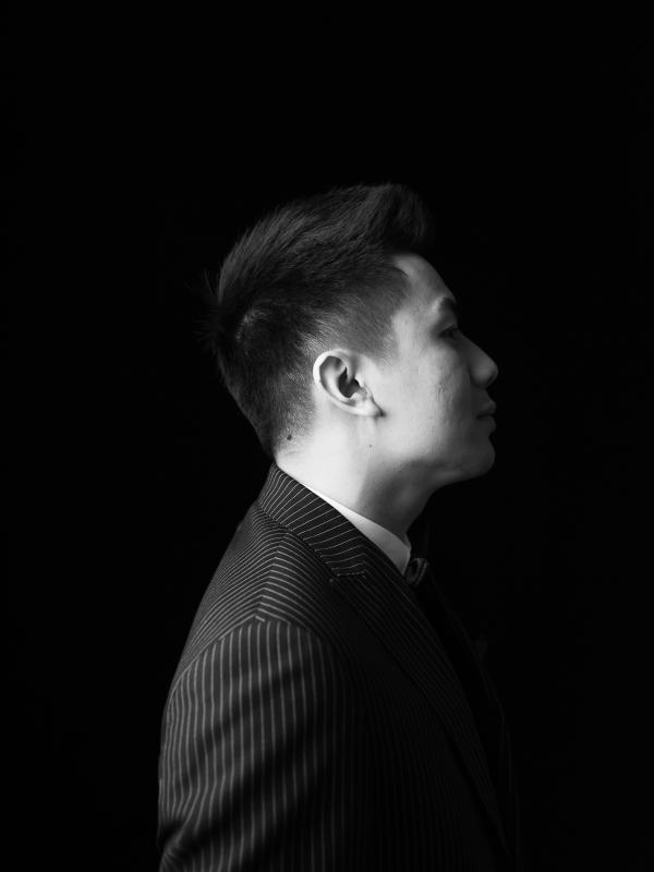
V.N. Trinh
· Assistant ProfessorVerifiedOhio State University · Arts and Sciences
Active 2006–2024
About
V.N. Trinh is an assistant professor in the Department of African American and African Studies and in the Center for Ethnic Studies at The Ohio State University. His academic research focuses on Black and Asian American radicals, their proto-abolitionist and anti-imperialist politics, and their encounters with state counterinsurgencies. He is currently drafting two books-in-progress: 'Burning All Illusions: Race and Rebellion in the City of Angels,' which is under contract with University of California Press and explores Los Angeles’ diverse, multi-racial left in relation to law-and-order policing; and 'Into the Belly of the Beast: Researchers, Radicals, and Refugees in Cold War California,' which investigates the relationship between the Silicon Valley military-industrial complex, opposition to the Vietnam-U.S. War by African American and Asian American communities, and the influx of Southeast Asian refugees to the San Francisco-Oakland Bay Area. Trinh has published shorter pieces through outlets such as Time, Washington Post, and Reappropriate. As an educator, he advocates for public schooling for all, including incarcerated individuals, and has been involved with the Yale Prison Education Initiative and the Ohio Prison Education Exchange Project, delivering courses to incarcerated Ohioans and coordinating related learning communities. Prior to his current position, he taught at Earlham College, Lafayette College, Southern Connecticut State University, and Yale University. He holds a PhD, two graduate degrees, and an undergraduate degree in history, all from Yale University and the University of California, Riverside, respectively.
Research topics
- Medicine
- Computer Science
- Nuclear medicine
- Physical medicine and rehabilitation
- Radiology
- Orthodontics
- Pathology
- Physical therapy
- Anatomy
- Traditional medicine
Selected publications
Current Problems in Diagnostic Radiology · 2024-05-03
articleThe American Journal of Sports Medicine · 2020 · 9 citations
1st authorCorresponding- Medicine
- Anatomy
- Nuclear medicine
BACKGROUND: Surgical treatment of symptomatic femoroacetabular impingement (FAI) and dysplasia requires careful characterization of acetabular morphology. The lateral center-edge angle (LCEA) is often used to assess lateral acetabular anatomy. Previous work has questioned the LCEA as a surrogate for acetabular contact/articular cartilage surface area because of the variable morphology of the lunate fossa. HYPOTHESIS: We hypothesized that weightbearing articular cartilage of the acetabulum would poorly correlate with LCEA secondary to significant variation in the size of the lunate fossa. STUDY DESIGN: Cohort study (Diagnosis); Level of evidence, 3. METHODS: < .05. RESULTS: = .88). Significant variation in surface area was observed within each group such that no patient in any group was outside of 2 SDs of the means of the other groups. When patients were categorized into quartiles established by the articular surface area for the entire population, 40% of patients with overcoverage were observed in the first or second quartile (lower area). CONCLUSION: Lateral acetabular undercoverage based on the LCEA (<25°) correlates with decreased acetabular surface area. Normal or increased acetabular coverage (LCEA, >25°), however, is not predictive of increased, normal, or decreased acetabular surface area.
Nonbiologic Injections in Sports Medicine
JBJS Reviews · 2020 · 10 citations
- Computer Science
- Traditional medicine
- Medicine
Nonbiologic medications (local anesthetics, corticosteroids, and nonsteroidal anti-inflammatory drugs) are commonly administered to athletes for analgesia after injury. However, the risks of nonbiologic injections often are overlooked simply because of their long-term market availability. A thorough understanding of the mechanism of action, the reported benefits, and the potential risks of nonbiologic medications is crucial prior to their use, especially in the treatment of young athletes. Sports medicine physicians and surgeons must be aware of the systemic and local effects of these medications to ensure an appropriate drug choice that minimizes side effects and avoids recently reported toxicity to myocytes, tenocytes, and chondrocytes.
Effect of Muscle Quality on Operative and Nonoperative Treatment of Rotator Cuff Tears
Orthopaedic Journal of Sports Medicine · 2019-08-01 · 17 citations
articleOpen accessBackground: Rotator cuff muscle atrophy and fatty infiltration are predictors of negative outcomes after rotator cuff repair. However, the impact of muscle degeneration on nonsurgical treatment is unknown. Hypothesis: Rotator cuff muscle atrophy and fatty infiltration will reduce the outcomes of operative repair while having a minimal effect on nonsurgical treatment. Additionally, in the setting of atrophy and fatty infiltration, surgical and nonsurgical treatment will produce equivalent outcomes. Study Design: Cohort study; Level of evidence, 2. Methods: Patients undergoing operative and nonoperative treatment of full-thickness rotator cuff tears were prospectively enrolled into a nonrandomized cohort study. Muscle quality was assessed on magnetic resonance imaging by use of the supraspinatus tangent sign, Warner atrophy, and Goutallier fatty infiltration classifications. Grading was performed by 2 independent observers who were blinded to patient treatment and outcomes. Normalized Western Ontario Rotator Cuff (WORC) index was the primary patient-reported outcome. Multivariate linear regression analysis was used to determine the impact of muscle quality on treatment outcomes. Results: The cohort consisted of 157 patients, 89 (57%) surgical and 68 (43%) nonsurgical, with a mean follow-up of 2.4 years (range, 1-5 years). Tangent sign had the best inter- and intrarater reliability, with kappa statistics of 0.81 and 0.86, respectively. Reliability for Warner atrophy was 0.69 to 0.76 and for Goutallier classification was 0.54 to 0.64. Overall, improvement in WORC scores was higher in the surgical group than the nonsurgical group (39.3 vs 21.2; P < .001). A positive tangent sign was the only independent predictor ( P < .01) of worse outcomes in the surgical group, accounting for an estimated 22-point lower improvement in WORC scores. Conclusion: A positive tangent sign was predictive of worse operative outcomes, resulting in equivalent improvements between surgical and nonsurgical treatment. The tangent sign is a reliable, prognostic indicator that clinicians can use when counseling patients on the optimal treatment of rotator cuff tears.
The American Journal of Sports Medicine · 2019-05-01 · 29 citations
article1st authorCorrespondingBACKGROUND: Traumatic anterior shoulder instability is a common condition affecting sports participation among young athletes. Clinical outcomes after surgical management may vary according to patient activity level and sport involvement. Overhead athletes may experience a higher rate of recurrent instability and difficulty returning to sport postoperatively with limited previous literature to guide treatment. PURPOSE: To report the clinical outcomes of patients undergoing primary arthroscopic anterior shoulder stabilization within the Multicenter Orthopaedic Outcomes Network (MOON) Shoulder Instability Consortium and to identify prognostic factors associated with successful return to sport at 2 years postoperatively. STUDY DESIGN: Case series; Level of evidence, 4. METHODS: Overhead athletes undergoing primary arthroscopic anterior shoulder stabilization as part of the MOON Shoulder Instability Consortium were identified for analysis. Primary outcomes included the rate of recurrent instability, defined as any patient reporting recurrent dislocation or reoperation attributed to persistent instability, and return to sport at 2 years postoperatively. Secondary outcomes included the Western Ontario Shoulder Instability Index and Kerlan-Jobe Orthopaedic Clinic Shoulder and Elbow questionnaire score. Univariate regression analysis was performed to identify patient and surgical factors predictive of return to sport at short-term follow-up. RESULTS: A total of 49 athletes were identified for inclusion. At 2-year follow-up, 31 (63%) athletes reported returning to sport. Of those returning to sport, 22 athletes (45% of the study population) were able to return to their previous levels of competition (nonrefereed, refereed, or professional) in at least 1 overhead sport. Two patients (4.1%) underwent revision stabilization, although 14 (28.6%) reported subjective apprehension or looseness. Age ( P = .87), sex ( P = .82), and baseline level of competition ( P = .37) were not predictive of return to sport. No difference in range of motion in all planes ( P > .05) and Western Ontario Shoulder Instability Index scores (78.0 vs 80.1, P = .73) was noted between those who reported returning to sport and those who did not. CONCLUSION: Primary arthroscopic anterior shoulder stabilization in overhead athletes is associated with a low rate of recurrent stabilization surgery. Return to overhead athletics at short-term follow-up is lower than that previously reported for the general athletic population.
Medial Patellofemoral Ligament Reconstruction Using a Partial Thickness Quadriceps Tendon Graft
Journal of the American Academy of Orthopaedic Surgeons · 2017-07-25
articleThis technique video demonstrates medial patellofemoral ligament (MPFL) reconstruction using a partial thickness quadriceps tendon graft. We’ve performed this procedure since 1998 and described the technique in 2005. We’ve been very pleased with the results. It’s a very simple technique in that you only have to detach and attach one side of the graft. The quadriceps tendon remains attached to the patella. It’s a flat, ribbon-like graft much like the native MPFL. It avoids patellar tunnels which are risks for fractures. We simply dissect and detach the graft proximally, rotate the graft, and attach it to the femur. In this manner, it lies along the course of the normal MPFL. Watch the video trailer: https://links.lww.com/JAAOS/A39.
Proximal Humerus Fracture-Dislocation with Laceration of the Axillary Artery: A Case Report.
PubMed · 2017-01-01 · 9 citations
articleOpen accessBACKGROUND: Proximal humerus fractures account for approximately 4-6% of all fractures. While the majority of these fractures are treated non-operatively, the amount of fracture displacement, concomitant injuries, and patient factors often result in the need for surgical stabilization. Although concomitant neurovascular injury in the setting of low-energy trauma is rare, injury to the surrounding neurovascular structures have previously been reported. METHODS: We report a case of a 79-year-old male who sustained a low energy fall resulting in a two-part fracture dislocation of the proximal humerus with an associated brachial plexopathy and axillary artery laceration. The patient underwent emergent open reduction internal fixation of his fracture in addition to an axillary-brachial artery bypass using a reverse-saphenous vein graft. The current article reports the presentation, management and prognosis of this rare injury. RESULTS: At 6 months, the patient demonstrated limited active shoulder abduction with no observed motor function at this elbow, wrist or hand. Radiographic follow up demonstrated a reduced glenohumeral joint with evidence avascular necrosis of the humeral head. CONCLUSION: Fracture-dislocations of the proximal humerus may be associated with significant neurovascular injury in the setting of low energy trauma. Despite early treatment, the prognosis of patients with this constellation of injuries is guarded.
The Anatomy of the Medial Patellofemoral Ligament
Orthopedics · 2017-02-27 · 12 citations
review1st authorCorrespondingRecurrent patellar dislocation is observed in many patients treated nonoperatively following primary dislocation. Injury to the medial patellofemoral ligament (MPFL) is reported in the majority of patients following dislocation. There is an increased interest in repair or reconstruction of the MPFL for patients experiencing recurrent instability. The femoral attachment of the MPFL is critical in determining graft behavior following reconstruction. The femoral attachment can be determined by referencing local anatomy, fluoroscopic imaging or on the basis of desired graft-length changes. This article reviews the anatomy of the MPFL, with a focus on its femoral insertion site as it pertains to anatomic, isometric, and anisometric reconstruction. [Orthopedics. 2017; 40(4):e583-e588.].
Sports · 2016-04-15 · 4 citations
articleOpen access1st authorAcute patellar dislocation remains a common injury in both adult and pediatric patients. Non-operative management has been advocated for patients without a history of recurrent instability. Although pathologic thresholds for consideration of operative management have previously been reported in adults, it is largely unknown in children. A retrospective review of all skeletally immature patients diagnosed with acute lateral patellar dislocation who had MRI imaging were included for analysis. An age-based control group was also identified. Six radiographic measurements were compared: lateral trochlear inclination (LTI), trochlear facet asymmetry (TFA), trochlear depth (TD), tibial tuberosity–trochlear groove (TT–TG), sulcus angle (SA) and patellar height ratio. A total of 178 patients were included for analysis (study: n = 108, control: n = 70). The mean age of patients in the study and control groups was 13.7 and 12.1 years respectively (p ≤ 0.001). Study group patients had significant differences in all radiographic measurements including a decreased LTI (p < 0.001), increased TFA (p < 0.001) and SA (p < 0.001). The mean trochlear depth was 3.4 mm and 5.6 mm for patients in the study and control groups respectively (p < 0.001). Study group patients had an increased patellar height ratio (p < 0.001) and TT–TG distance (p < 0.001). Morphologic abnormalities may predispose skeletally immature patients to an increased risk of acute lateral patellar instability.
Computational analysis of factors contributing to patellar dislocation
Journal of Orthopaedic Research® · 2015-09-02 · 88 citations
articleOpen accessTreatment for an initial incidence of patellar dislocation is usually conservative management; however, almost half of patients experience a subsequent, or multiple, dislocation(s). Patients often undergo multiple procedures which fail to treat the underlying anatomic abnormalities. The objective of this study was to evaluate interactions between key predisposing anatomic factors to patellar dislocation and identify combinations of abnormal factors which increase the risk of recurrent lateral dislocation. Four factors associated with lateral patellar dislocation were identified (sulcus angle, Insall-Salvati ratio, tibial tubercle-trochlear groove distance, and femoral anteversion). A finite element model of the patellofemoral joint was developed and parameterized so that a value for each factor could be applied and the model geometry/alignment would be modified accordingly. 100 combinations of the four factors were generated in separate computational simulations and resulting kinematics and forces of the patellofemoral joint were recorded. Sulcus angle was the most impactful factor on constraint. Multiple abnormal factors were generally required to produce the extremes of patellar alignment observed in this analysis. Understanding the underlying anatomic factors, and their effect on joint mechanics, for patients with recurrent lateral patellar dislocation will aid in determining optimal treatment pathways on a patient-specific basis.
Frequent coauthors
- 6 shared
Benjamin R. Pulley
- 6 shared
Asheesh Bedi
- 6 shared
David C. Flanigan
The Ohio State University Wexner Medical Center
- 6 shared
Jason R. Ferrel
University of Utah
- 6 shared
Joshua D. Harris
Methodist Hospital
- 5 shared
T. Ty Fowler
Mount Carmel Health
- 5 shared
Jared C. Bentley
Steadman Clinic
- 4 shared
Michael Leunig
Schulthess-Klinik
- Resume-aware match score
- Save to shortlist
- AI-drafted outreach
See your match with V.N. Trinh
PhdFit ranks faculty by your research interests, methods, and publications — grounded in their actual work, not templates.
- Free to start
- No credit card
- 30-second signup